Category: Myoclonus/Tics/Stereotypies
Objective: We sought to develop a standardised, computationally tractable approach to the analysis of electrophysiology in orthostatic myoclonus by developing novel definition criteria for myoclonic bursts, allowing automated burst identification for further analysis.
Background: Orthostatic myoclonus refers to irregular, myoclonic electromyography (EMG) bursts triggered by standing and is the most common cause of unsteadiness on standing.[1] There is limited literature on its electrophysiological characteristics.[2]
Method: 15 second epochs of surface EMG recordings from the lower limb muscles (Table 1) during stance of ten subjects (age 46-93 years, 4 females and 6 males) with orthostatic myoclonus were analysed using MATLAB. Raw signals were 50-200 Hz band-passed filtered, rectified and enveloped using a moving 50 ms root mean square (RMS) window [3] to minimise noise and signal fluctuation. Three different burst criteria were evaluated: amplitude ≥ 25%, 50% or 2 SD above the RMS background with duration ≥ 20 ms. Criteria demonstrating greatest concordance with expert clinician-marked bursts were then applied to assess other features. Synchronicity was quantified by the percentage of bursts in muscle pairs with onset ≤ 15 ms of each other.[2] Rhythmicity was defined as having at least one frequency peak ≥ 3 times the power of neighbouring +/- 1 Hz frequencies on the Fourier transform.
Results: Amplitude ≥ 25% background and duration ≥ 20 ms criteria exhibited greatest concordance with clinician markings and was used for subsequent analysis. Mean burst duration in all muscles was 50-100 ms (Table 1). Greatest synchronicity was seen between homologous lower leg muscles, maximal in tibialis anterior (Table 2). Rhythmicity in at least one muscle was seen in 4/10 subjects with 3/6 instances occurring in biceps femoris (2-10 Hz), 2/6 in quadriceps (1.5 Hz) and 1/6 in tibialis anterior (9.5 Hz).
Conclusion: Our novel computational method for EMG analysis successfully characterised key electrophysiological features of orthostatic myoclonus, consistent with published data.[2] Our method has the advantage of being standardised, efficient and scalable compared to manual analysis by clinicians and application to a larger cohort for more extensive analysis is planned.
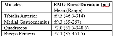
Table 1: EMG burst duration.
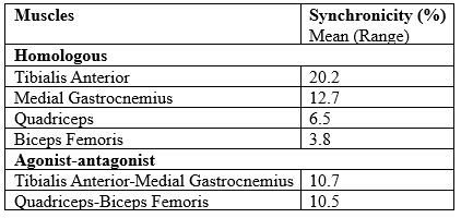
Table 2: Synchronicity of EMG bursts.
References: 1. Gasca-Salas C, Arcocha J, Artieda J, Pastor P. Orthostatic myoclonus: an underrecognized cause of unsteadiness? Parkinsonism Relat Disord 2013;19:1013–1017.
2. Glass GA, Ahlskog JE, Matsumoto JY. Orthostatic myoclonus: a contributor to gait decline in selected elderly. Neurology 2007;68(21):1826-30.
3. Carvalho CR, Fernández JM, del-Ama AJ, Oliveira Barroso F, & Moreno JC. Review of electromyography onset detection methods for real-time control of robotic exoskeletons. J. Neuroengineering Rehabil 2023;20(1):1–141.
To cite this abstract in AMA style:
N. Jeyakumar, S. Nagaratnam, G. Jeyakumar, P. Becker, F. Chang, V. Fung. A Standardised, Semi-automated Computational Approach to the Analysis of Electrophysiology in Orthostatic Myoclonus [abstract]. Mov Disord. 2025; 40 (suppl 1). https://www.mdsabstracts.org/abstract/a-standardised-semi-automated-computational-approach-to-the-analysis-of-electrophysiology-in-orthostatic-myoclonus/. Accessed July 7, 2026.« Back to 2025 International Congress
MDS Abstracts - https://www.mdsabstracts.org/abstract/a-standardised-semi-automated-computational-approach-to-the-analysis-of-electrophysiology-in-orthostatic-myoclonus/