Objective: To assess structural brain alterations in patients with cervical dystonia (CD) and focal upper limb dystonia (ULD) relative to healthy controls, using MRI exploratory analysis and region of interest techniques.
Background: The striatum is integral to motor control and is associated with dystonia and other movement disorders. A novel diffusion-weighted imaging (DWI) technique now distinguishes between matrix-like and striosome-like compartments of the striatum in vivo through connectivity-based parcellation. Characterizing these compartments in focal dystonia subtypes may offer new insights into the disease’s pathophysiology.
Method: The study comprised 91 participants: 29 with ULD, 25 with CD, and 37 healthy controls, all imaged on the same Siemens 3.0T scanner using T1 and DWI. Data processing was conducted using FreeSurfer and FSL, with analysis through whole-brain and region of interest approaches using permutation-based statistics, accounting for age, sex, and multiple comparisons.
Results: Dystonia patients showed significant reductions in the left caudate and matrix-like voxel volumes compared to controls. Severity of dystonia was inversely correlated with bilateral putamen volumes. Specifically, CD patients had reduced volumes in the right thalamus and left caudate compared to controls, along with significant increases in radial diffusivity within white matter tracts. ULD patients exhibited reduced matrix-like voxel volumes in the left striatum, with no significant changes in other subcortical structures. Notably, CD patients also showed decreased volume in the right thalamus compared to ULD and HC. No significant differences in cortical thickness, area, or volume were observed between the groups.
Conclusion: ThisThis study unveils specific structural brain alterations in CD and ULD, with significant involvement of the striatum and its associated white matter pathways. Notably, while the cortical architecture was preserved across subtypes of focal dystonia, our findings highlight distinct microstructural changes in the basal ganglia that correlate with the clinical severity and manifestations observed in CD and ULD. These insights deepen our understanding of the neurobiological mechanisms that differentiate these dystonia subtypes, potentially guiding targeted therapeutic strategies.
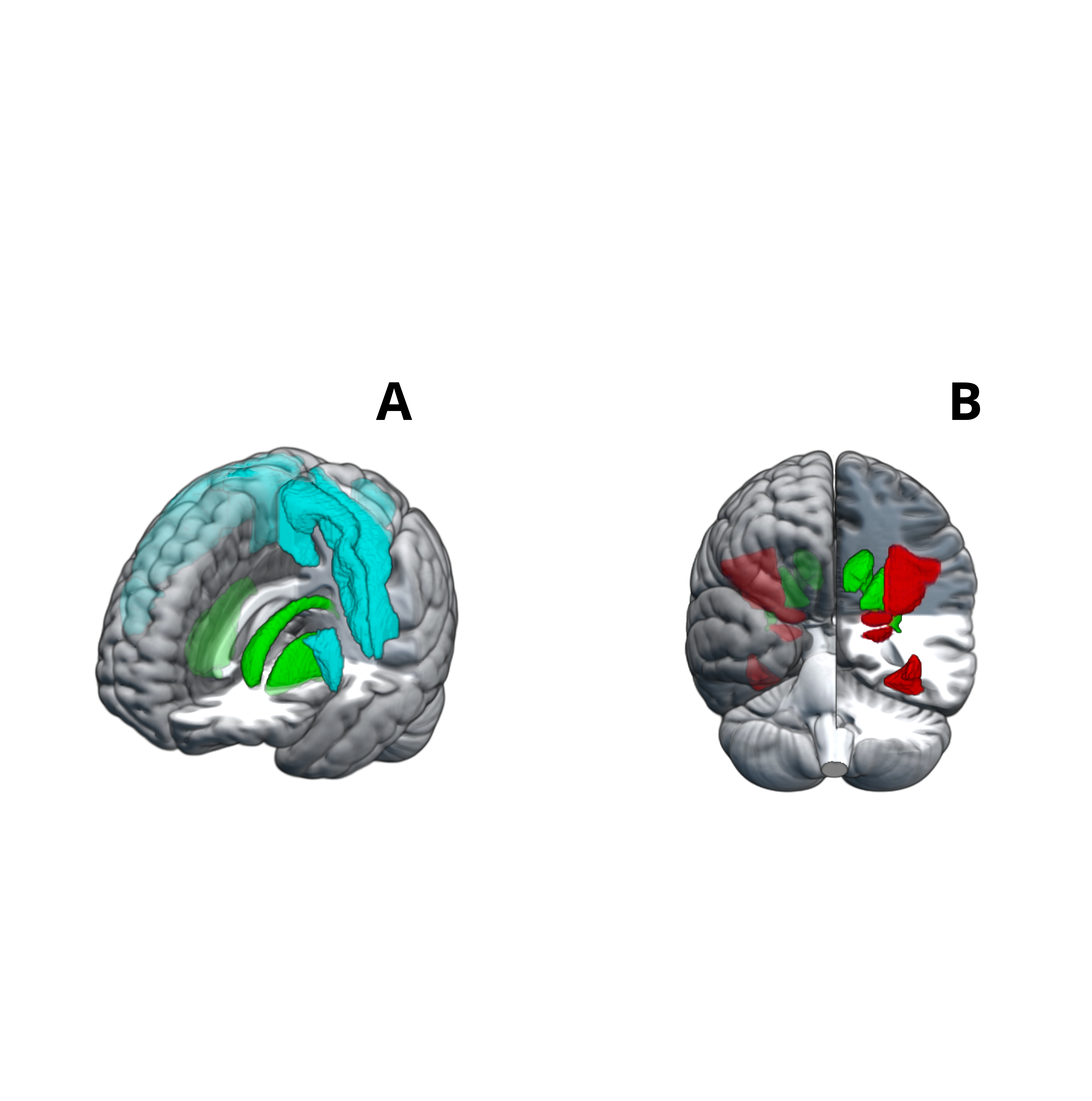
Connectivity-based parcellation of striatum.
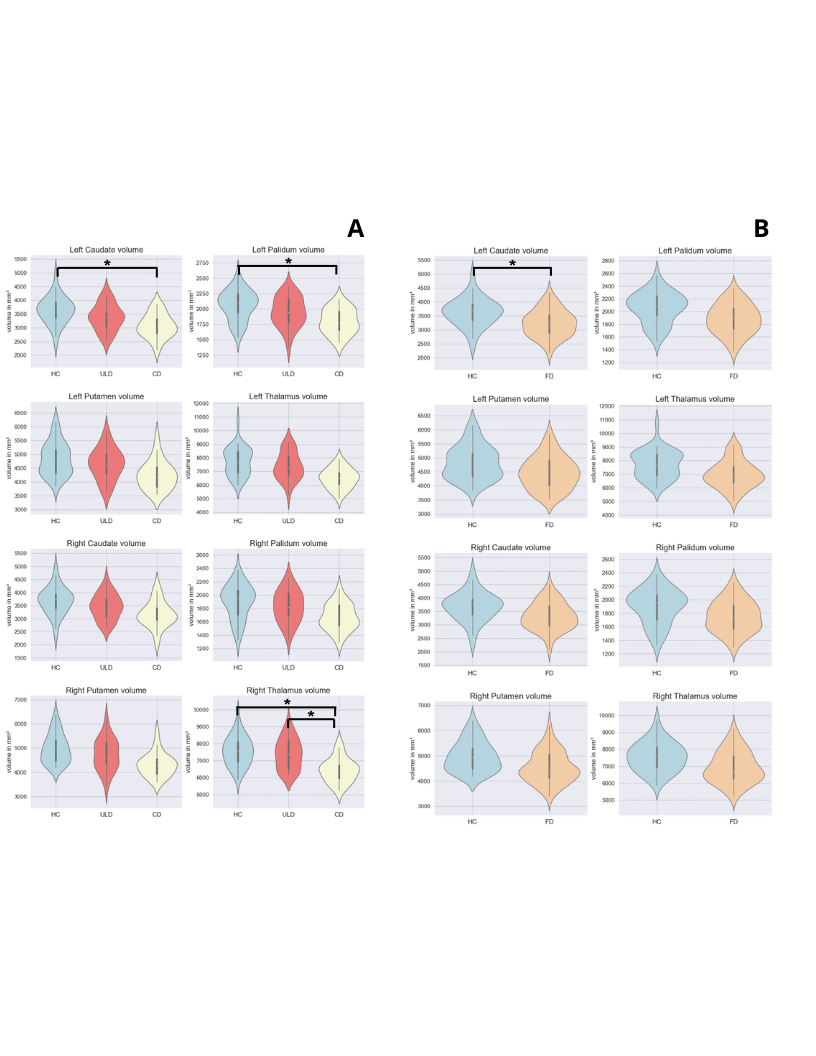
Bsal ganglia volume
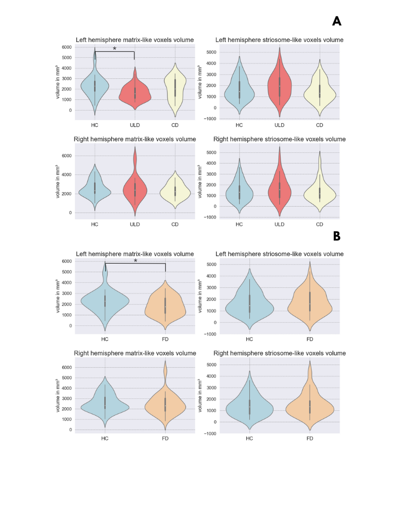
Striosome- and Matrix-like voxel volume
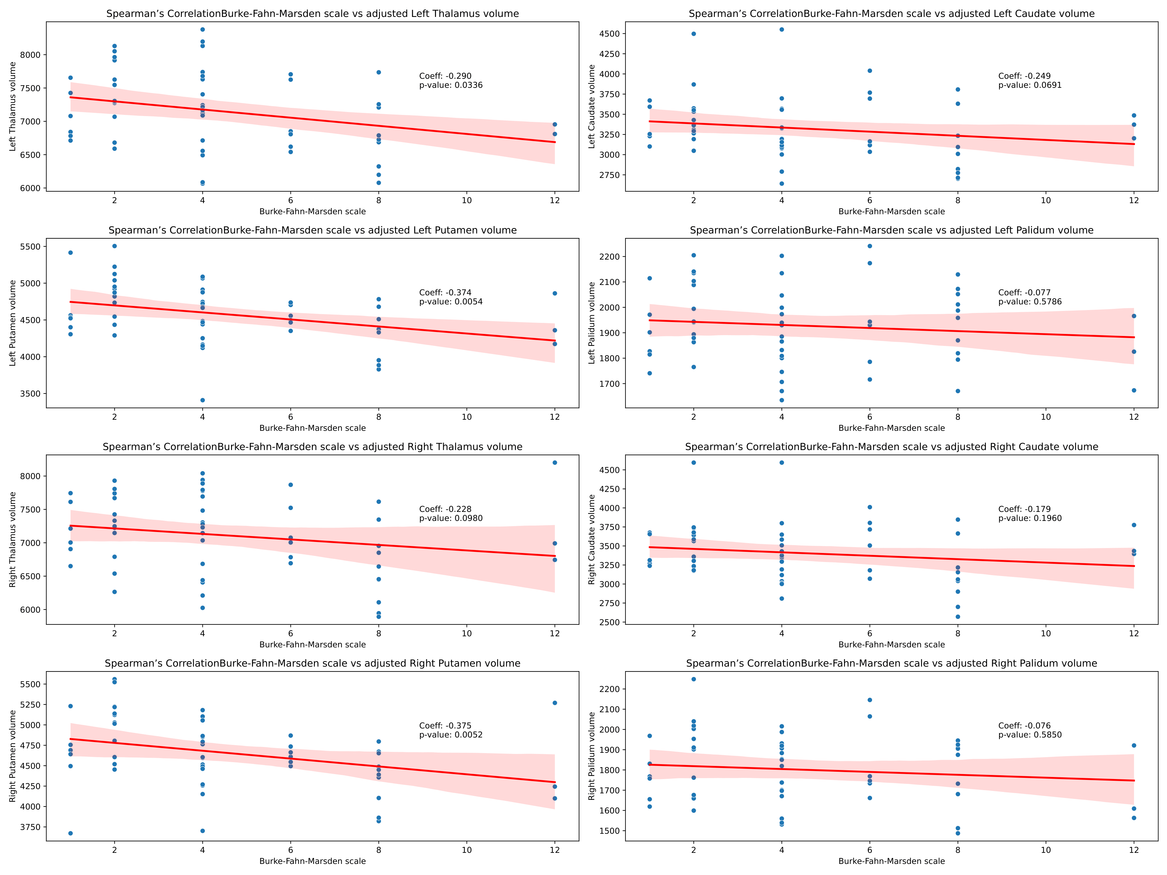
Basal ganglia volume and dystonia severity
References: Waugh JL, Hassan AAO, Kuster JK, Levenstein JM, Warfield SK, Makris N, et al. An MRI method for parcellating the human striatum into matrix and striosome compartments in vivo. Neuroimage. 2022;246:118714.
Winkler AM, Ridgway GR, Webster MA, Smith SM, Nichols TE. Permutation inference for the general linear model. NeuroImage. 2014;92:381–97.
To cite this abstract in AMA style:
J. de Paiva, D. de Faria, V. Borges, S. Silva, H. de Ferraz, P. Aguiar. White Matter and Subcortical Alterations in Focal Dystonia: A MRI Study of Subtype-Specific Brain Changes [abstract]. Mov Disord. 2025; 40 (suppl 1). https://www.mdsabstracts.org/abstract/white-matter-and-subcortical-alterations-in-focal-dystonia-a-mri-study-of-subtype-specific-brain-changes/. Accessed July 5, 2026.« Back to 2025 International Congress
MDS Abstracts - https://www.mdsabstracts.org/abstract/white-matter-and-subcortical-alterations-in-focal-dystonia-a-mri-study-of-subtype-specific-brain-changes/