Category: Parkinson's disease: Neuroimaging
Objective: This study aims to examine iron deposition in striatum and substantia nigra pars compacta (SNc) functionally segmented in patients with isolated rapid eye movement sleep behavior disorder (iRBD) and its relationship with clinical features and neuropsychological performance.
Background: iRBD is recognized as a prodromal stage of synucleinopathies [1]. Iron deposition in basal ganglia (BG) is thought to be crucial in the physiopathology of neurodegenerative diseases, promotoing neural death and inflammation [2]. R2*-weighted imaging can quantify iron content by employing magnetic susceptibility effects [3] and R2* increases have been identified in iRBD [4], remarking its potential as an early imaging biomarker. However, the relationship between iron deposition in nigrostriatal system functional regions of SNc and BG, and its relationship with clinical and neuropsychological features has been poorly studied.
Method: Thirty-three iRBD and 24 healthy controls (HC) participants underwent a neuropsychological assessment. The R2* maps were obtained by fitting the exponential decay voxel by voxel. Segmented striatum [5] and SNc [6] were obtained with functional connectivity atlases. Between-group differences in R2* values and partial correlations between striatum and clinical features were computed adjusting for sex. All the results were corrected by false discovery rate (p<0.05).
Results: Patients with iRBD presented higher R2* values in limbic (mean HC=0.022, mean iRBD=0.025) and sensorimotor striatum (mean HC=0.0054, mean iRBD=0.0053) compared to HC but showed no differences in SNc [Figure 1]. iRBD also presented lower performance in Mini-Mental State Examination, University of Pennsylvania Smell Identification Test, Digit span forward, Symbol Digit Modalities Test (SDMT), Stroop Words, and neuropsychiatric inventory [Table 1 and 2]. The sensorimotor striatum correlated negatively with SDMT in iRBD (r=-2.97, p=0.006) [Figure 2].
Conclusion: Iron deposition in limbic and sensorimotor striatum is present in prodromic stages of synucleinopathies. Moreover, sensorimotor striatum iron content reflects speed processing decay in iRBD, indicating an association between iron deposition and clinical features in these patients. Overall, these results indicate the potential of noninvasive iron estimates imaging as an early biomarker in synucleopathies.
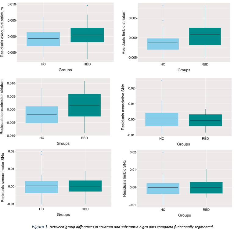
Figure 1
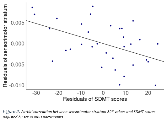
Figure 2
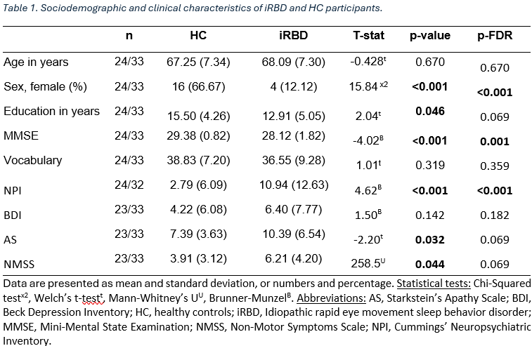
Table 1
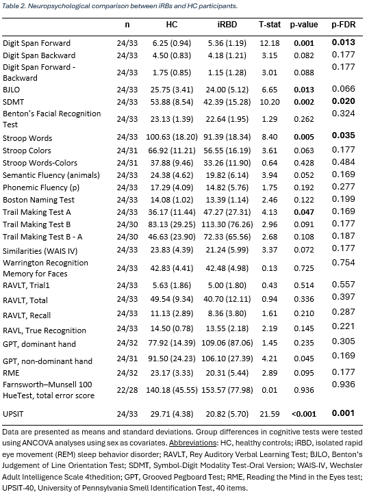
Table 2
References: [1] Iranzo A, Santamaria J, Tolosa E. Idiopathic rapid eye movement sleep behaviour disorder: diagnosis, management, and the need for neuroprotective interventions. Lancet Neurol. 2016;15(4):405-419. doi: 10.1016/S1474-4422(16)00057-0.
[2] Zeng, W., Cai, J., Zhang, L., & Peng, Q. (2024). Iron Deposition in Parkinson’s Disease: A Mini-Review. Cellular and molecular neurobiology, 44(1), 26. https://doi.org/10.1007/s10571-024-01459-4
[3] Langkammer, C., Krebs, N., Goessler, W., Scheurer, E., Ebner, F., Yen, K., Fazekas, F., & Ropele, S. (2010). Quantitative MR imaging of brain iron: a postmortem validation study. Radiology, 257(2), 455–462. https://doi.org/10.1148/radiol.10100495
[4] Nepozitek, J., Varga, Z., Dostalova, S., Perinova, P., Keller, J., Robinson, S., Ibarburu, V., Prihodova, I., Bezdicek, O., Ruzicka, E., Sonka, K., & Dusek, P. (2023). Magnetic susceptibility changes in the brainstem reflect REM sleep without atonia severity in isolated REM sleep behavior disorder. NPJ Parkinson’s disease, 9(1), 112. https://doi.org/10.1038/s41531-023-00557-2
[5] Tziortzi et al. Imaging dopamine receptors in humans with [11C]-(+)-PHNO: dissection of D3 signal and anatomy. NeuroImage 54: 264-77 (2011) https://fsl.fmrib.ox.ac.uk/fsl/fslwiki/Atlases/striatumstruc
[6] Biondetti, E., Santin, M. D., Valabrègue, R., Mangone, G., Gaurav, R., Pyatigorskaya, N., Hutchison, M., Yahia-Cherif, L., Villain, N., Habert, M. O., Arnulf, I., Leu-Semenescu, S., Dodet, P., Vila, M., Corvol, J. C., Vidailhet, M., & Lehéricy, S. (2021). The spatiotemporal changes in dopamine, neuromelanin and iron characterizing Parkinson’s disease. Brain: a journal of neurology, 144(10), 3114–3125. https://doi.org/10.1093/brain/awab191
To cite this abstract in AMA style:
C. Garcia-Vicente, I. Roura, J. Pardo, C. Martín-Barceló, C. Falcon, R. Sala-Llonch, N. Bargalló, M. Serradell, C. Gaig, G. Mayà, A. Montini, C. Junqué, A. Iranzo, B. Segura. Sensory-motor striatum iron accumulation in patients with isolated rapid eye movement sleep behavior disorder and its correlation with processing speed performance [abstract]. Mov Disord. 2025; 40 (suppl 1). https://www.mdsabstracts.org/abstract/sensory-motor-striatum-iron-accumulation-in-patients-with-isolated-rapid-eye-movement-sleep-behavior-disorder-and-its-correlation-with-processing-speed-performance/. Accessed July 7, 2026.« Back to 2025 International Congress
MDS Abstracts - https://www.mdsabstracts.org/abstract/sensory-motor-striatum-iron-accumulation-in-patients-with-isolated-rapid-eye-movement-sleep-behavior-disorder-and-its-correlation-with-processing-speed-performance/