Category: Parkinson's disease: Neuroimaging
Objective: This prospective cohort study longitudinally investigated hippocampal subfield shape deformation and atrophy in early-stage Parkinson’s Disease (PD), using spherical harmonic-based shape analysis.
Background: Hippocampal subfields are important player in cognitive function in PD. Current study focuses on volumetric analysis describing atrophy, where limited research established on shape analysis of the hippocampal subfield morphometry.
Method: A cohort of early PD (n = 146; disease duration = 14.9 ± 9.5 months; Gender = 44% Female) and matched healthy controls (HC) (n = 59; Gender = 56% Female) underwent MRI and cognitive assessments with follow-up after two years [table1]. Hippocampal subfields were segmented using FreeSurfer, and shape deformations were analysed via SPHARM-PDM module.
Results: Shape analysis identified selective morphological changes in hippocampal subfields (CA1, CA3, subiculum, and fimbria) at baseline in early PD patients compared to HCs, despite no volumetric differences [figure1]. Longitudinally, atrophy in the right whole hippocampus, and selective hippocampal subfields (right CA1, and left fimbria) were present. In the PD-Mild Cognitive Impairment (PD-MCI) subgroup (n = 27), localised shape deformations in CA1, CA3, GC-ML-DG, and fimbria were observed at baseline when compared to PD-Normal Cognition (PD-NC) (n = 84) [figure1]. Longitudinal subfield left fimbria atrophy emerged as a sensitive biomarker predicting cognitive deterioration, especially in visuospatial and memory domains.
Conclusion: Shape analysis provides superior sensitivity over volumetric analysis for detecting neurodegeneration that precedes gross volumetric atrophy in de-novo PD. This study highlights the potential of hippocampal subfield shape analysis for early detection of cognitive decline in PD. One of the subfields, left fimbria, arises as a promising biomarker for predicting cognitive decline in PD and guiding early targeted interventions.
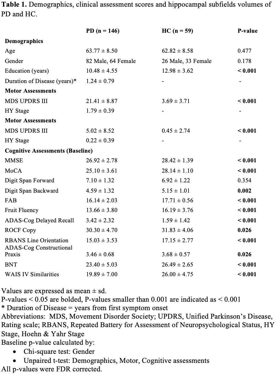
Table 1
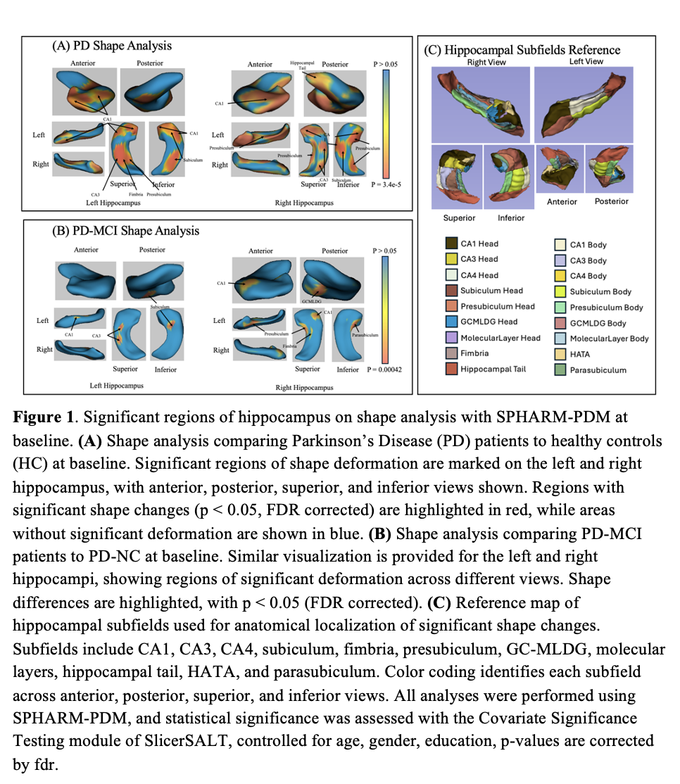
Figure 1
To cite this abstract in AMA style:
T. Wu, S. Ng, T. Teo, X. Choi, D. Heng, S. Neo, Z. Xu, K. Tay, W. Au, E. Tan, L. Chan, L. Tan, T. Welton. Longitudinal MRI Study of Hippocampal Subfields Morphometry in Early Parkinson’s Disease [abstract]. Mov Disord. 2025; 40 (suppl 1). https://www.mdsabstracts.org/abstract/longitudinal-mri-study-of-hippocampal-subfields-morphometry-in-early-parkinsons-disease/. Accessed July 7, 2026.« Back to 2025 International Congress
MDS Abstracts - https://www.mdsabstracts.org/abstract/longitudinal-mri-study-of-hippocampal-subfields-morphometry-in-early-parkinsons-disease/