Category: Parkinson's disease: Neuroimaging
Objective: We assessed patterns of dopamine (DA) release and striatal activation induced by behavioural tasks involving corticostriatal loops in Parkinson’s disease (PD) and healthy control (HC) subjects using positron emission tomography (PET) with 11C-raclopride.
Background: DA striatal denervation is clinically associated with cardinal motor features of PD. We hypothesized that DA depletion would lead to a loss of discrete somatotopic representation within the striatum and might disrupt the functional segregation of motor, associative and limbic circuits.
Method: Thirteen early-stage PD and 15 gender- and aged-matched HC subjects underwent 4 PET/MRI scans, each following a single bolus infusion of 11C-raclopride (550 MBq). Activation tasks included: (i) finger tapping, (ii) foot tapping, (iii) a card-sorting task; (iv) a card task with a monetary reward. PET data were analyzed with RSD-Hybrid-IMRTM to estimate DA release. Principal component analysis (PCA) was used to assess group differences in spatial patterns of DA quantified by the RSD-Hybrid-IMRTM-derived parameter beta (1).
Results: During finger tapping, PD subjects exhibited greater activation in the ipsilateral caudate and ventral striatum, while activation in the contralateral caudate and bilateral dorsal putamen was lower compared to HC. In foot tapping, PD subjects showed higher DA release in the bilateral caudate and ventral striatum, but reduced release in the bilateral dorsal putamen compared to HC (Figure 1). When comparing the foot tapping vs finger tapping, HC showed relatively greater DA release in the contralateral ventral caudate, associated with relatively reduced release in the dorsal putamen (Figure 2); however, no such differences were observed in PD. In both reward and cognitive tasks, there was relatively greater activation of bilateral caudate and ventral striatum and reduced activation of more affected putamen in PD compared to HC. Across all tasks, there was significant overlap of DA release in the less affected striatum of PD, while in HC, overlap was minimal.
Conclusion: Compared with HC, PD subjects exhibited distinct activation patterns, with predominant activation in large ipsilateral clusters during tapping. In HC, functional loops are typically segregated, with clear anatomical distinctions between striatal regions associated with specific functions. However, in PD, these distinctions appear blurred, suggesting a loss of anatomical segregation of DA release.
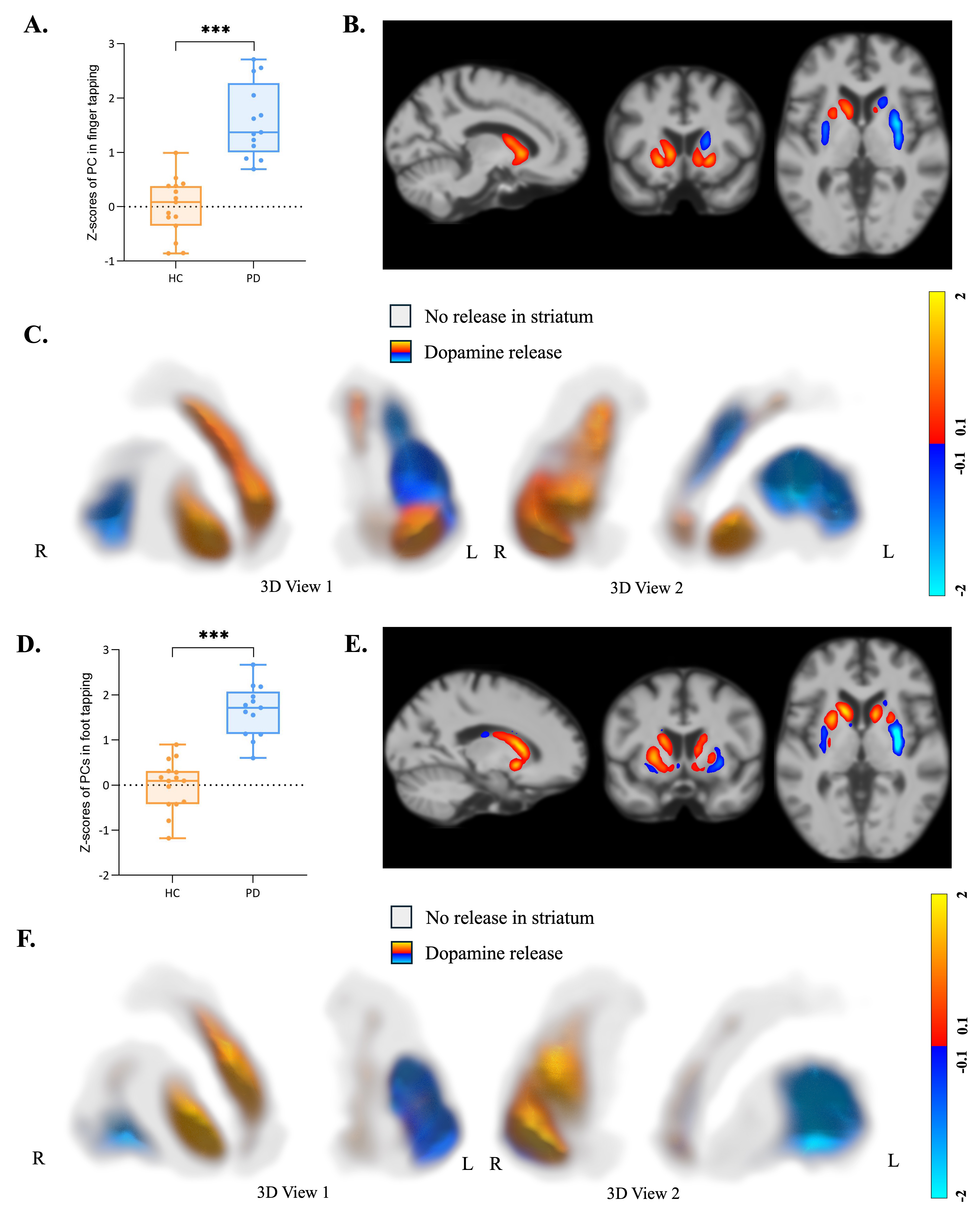
Figure 1
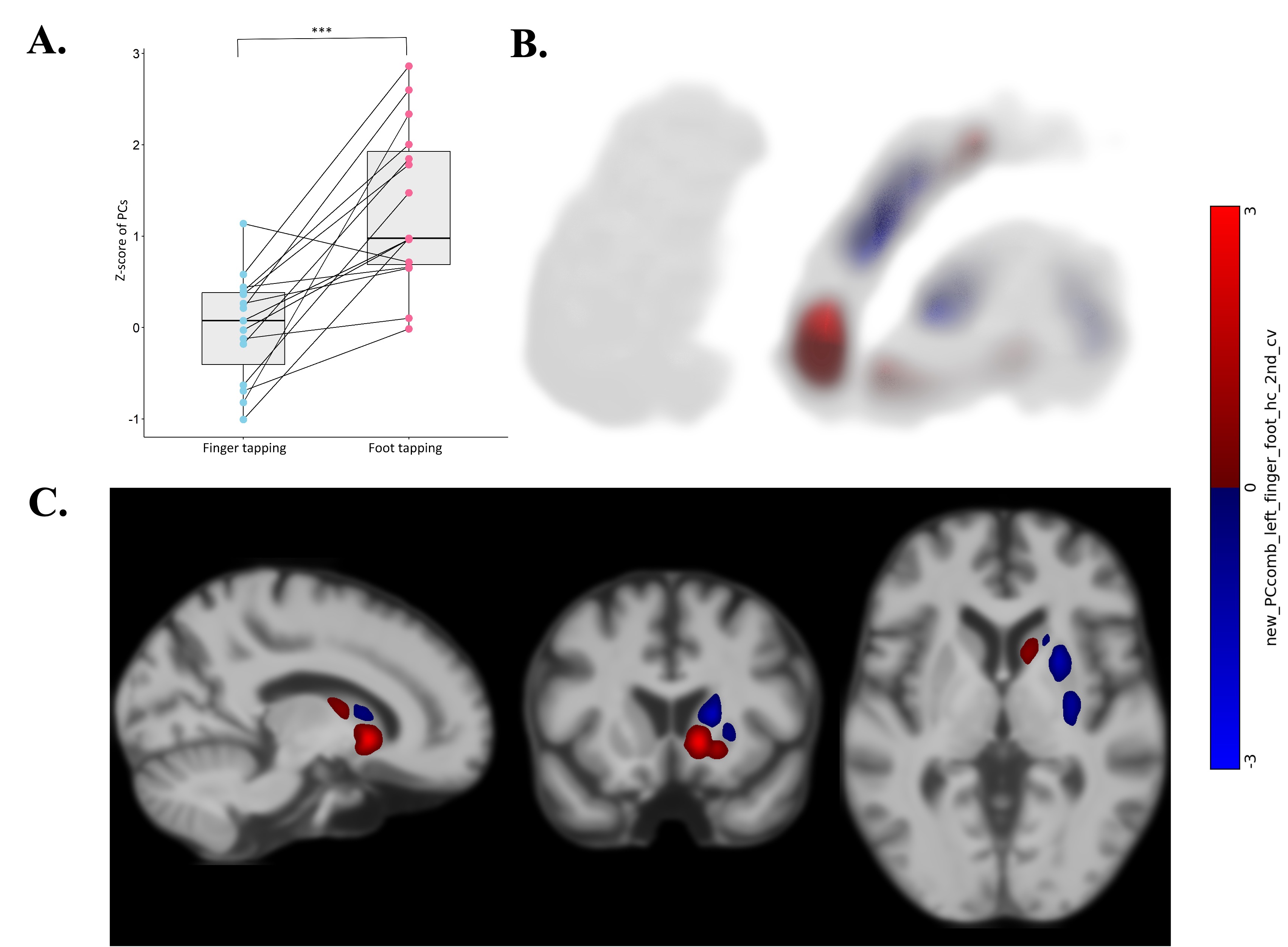
Figure 2
References: [1] Bevington CW, Hanania JU, Ferraresso G, Cheng JK, Pavel A, Su D, et al. Novel voxelwise residual analysis of [(11)C]raclopride PET data improves detection of low-amplitude dopamine release. J Cereb Blood Flow Metab. 2024;44(5):757-71.
To cite this abstract in AMA style:
DN. Su, J. Hanania, A. Pavel, M. Matarazzo, J. Mckenzie, J. Cheng, N. Vafai, S. Dhaliwal, V. Sossi, A. Stoessl. Functional Segregation in Parkinson’s Disease [abstract]. Mov Disord. 2025; 40 (suppl 1). https://www.mdsabstracts.org/abstract/functional-segregation-in-parkinsons-disease/. Accessed July 7, 2026.« Back to 2025 International Congress
MDS Abstracts - https://www.mdsabstracts.org/abstract/functional-segregation-in-parkinsons-disease/