Category: Parkinson's disease: Neuroimaging
Objective: To assess whether advanced diffusion magnetic resonance imaging (dMRI) techniques help to understand the outcome of Deep brain stimulation (DBS) surgery on Unified Parkinson’s Disease Rating Scale (UPDRS) and neuropsychological measures.
Background: DBS is a surgical option for patients with PD patients with fluctuations in motor function2, 3 but outcomes are variable4. Advanced neuroimaging methods5 and connectomics approach6 could inform targeting and post-operative programming adjustment,. Connectomics derived from diffusion MRI (dMRI) suffer from fiber crossings at clinical resolution of acquisition7,8. We hypothesized that white matter (WM) organizational properties derived using advanced dMRI techniques such as fiber density and cross section (FDC) could help in understanding DBS outcomes.
Method: We acquired conventional single-shell dMRI data from 31 patients with PD who underwent unilateral subthalamic nucleus (STN) DBS in one brain hemisphere9. TR=9.6s, TE=101ms, resolution=2x2x2.75mm, number of diffusion encoding directions=90, number of interleaved b0s=12, b-values=1000s/mm2, slice encoding direction=A>>P; along with T1-weighted MRI with 0.9×0.9x1mm resolution. We performed denoising10, Gibbs deringing11, and eddy current correction and computed voxel-level FDC on all participants using MrTrix312. A population-based template based on our cohort was generated to perform correlational analysis between voxel-level FDC, UPDRS, and cognitive measures. The statistical analysis was conducted separately for left and right hemispheric candidates for DBS to understand lateralization effects. Results were signficant at uncorrected p-value of 0.001.
Results: For left hemisphere DBS implantation, we found significant positive correlations with FDC and both UPDRS (fig. 1) and verbal fluency (fig. 2) at baseline; and a positive association with FDC and cognitive switching (fig. 3) change from baseline. For right hemisphere DBS implantation, we found significant positive correlations with FDC and UPDRS, verbal fluency, and cognitive inhibition scores; and a negative association between FDC and UPDRS change from baseline.
Conclusion: Beyond single-tensor dMRI-analysis that accounts for fiber crossings shows promise for understanding DBS outcomes and should be further examined in independent retrospective and prospective cohorts.
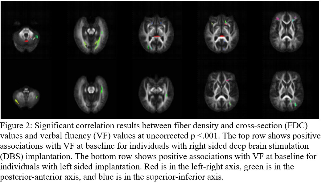
Figure 2
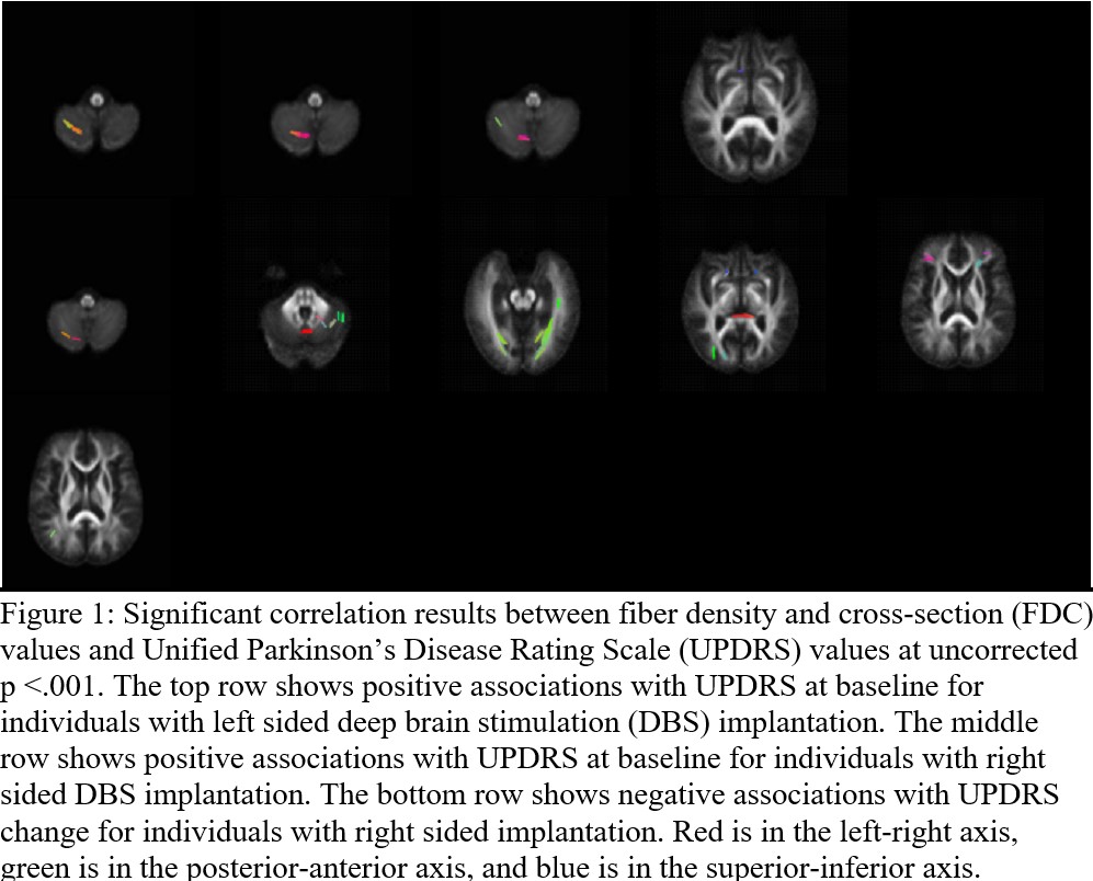
Figure 1
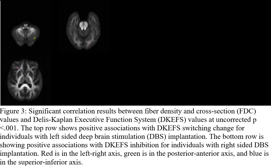
Figure 3
References: 1. Stocchi F, Bravi D, Emmi A, Antonini A. Parkinson disease therapy: current strategies and future research priorities. Nature Reviews Neurology 2024.
2. Deuschl G, Schade-Brittinger C, Krack P, et al. A randomized trial of deep-brain stimulation for Parkinson’s disease. N Engl J Med 2006;355:896-908.
3. Kleiner-Fisman G, Herzog J, Fisman DN, et al. Subthalamic nucleus deep brain stimulation: summary and meta-analysis of outcomes. Mov Disord 2006;21 Suppl 14:S290-304.
4. Bratsos S, Karponis D, Saleh SN. Efficacy and Safety of Deep Brain Stimulation in the Treatment of Parkinson’s Disease: A Systematic Review and Meta-analysis of Randomized Controlled Trials. Cureus 2018;10:e3474.
5. Peng S, Dhawan V, Eidelberg D, Ma Y. Neuroimaging evaluation of deep brain stimulation in the treatment of representative neurodegenerative and neuropsychiatric disorders. Bioelectronic Medicine 2021;7:4.
6. Horn A, Fox MD. Opportunities of connectomic neuromodulation. NeuroImage 2020;221:117180.
7. Mishra V, Guo X, Delgado MR, Huang H. Toward tract-specific fractional anisotropy (TSFA) at crossing-fiber regions with clinical diffusion MRI. Magnetic resonance in medicine : official journal of the Society of Magnetic Resonance in Medicine / Society of Magnetic Resonance in Medicine 2014;00:1-12.
8. Jeurissen, B., Leemans, A., Tournier, J. D., Jones, D. K., & Sijbers, J. (2013). Investigating the prevalence of complex fiber configurations in white matter tissue with diffusion magnetic resonance imaging. Human brain mapping, 34(11), 2747–2766. https://doi.org/10.1002/hbm.22099
9. Del Bene, V. A., Martin, R. C., Brinkerhoff, S. A., Olson, J. W., Nelson, M. J., Marotta, D., Gonzalez, C. L., Mills, K. A., Kamath, V., Bentley, J. N., Guthrie, B. L., Knight, R. T., & Walker, H. C. (2023). Differential cognitive effects of unilateral left and right subthalamic nucleus deep brain stimulation for Parkinson disease. medRxiv : the preprint server for health sciences, 2023.02.27.23286478. https://doi.org/10.1101/2023.02.27.23286478
10. Veraart J, Novikov DS, Christiaens D, Ades-Aron B, Sijbers J, Fieremans E. Denoising of diffusion MRI using random matrix theory. Neuroimage 2016;142:394-406.
11. Kellner E, Dhital B, Kiselev VG, Reisert M. Gibbs-ringing artifact removal based on local subvoxel-shifts. Magn Reson Med 2016;76:1574-1581
12. Tournier JD, Smith R, Raffelt D, et al. MRtrix3: A fast, flexible and open software framework for medical image processing and visualisation. Neuroimage 2019;202:116137.
To cite this abstract in AMA style:
I. Campbell, V. Del Bene, R. Martin, C. Gonzalez, S. Brinkerhoff, M. Wade, B. Hill, H. Walker, V. Mishra. Subthalamic nucleus DBS outcomes correlate with white matter tractography in Parkinson’s Disease [abstract]. Mov Disord. 2025; 40 (suppl 1). https://www.mdsabstracts.org/abstract/subthalamic-nucleus-dbs-outcomes-correlate-with-white-matter-tractography-in-parkinsons-disease/. Accessed July 7, 2026.« Back to 2025 International Congress
MDS Abstracts - https://www.mdsabstracts.org/abstract/subthalamic-nucleus-dbs-outcomes-correlate-with-white-matter-tractography-in-parkinsons-disease/