Category: Parkinson's disease: Neuroimaging
Objective: To define the subtypes of Parkinson’s disease (PD) based on the spatiotemporal progression patterns of cerebral hypoperfusion.
Background: Early-phase 18F-FP-CIT PET images, which are available in a single PET session when diagnosing PD, have proven useful in exploring the cerebral perfusion changes in response to PD-related neurodegeneration [1-4], and may potentially uncover the neural basis of the clinical heterogeneity of PD.
Method: A total of 655 patients with PD and 48 healthy controls who underwent dual-phase 18F-FP-CIT PET scans were retrospectively enrolled. After quantifying early-phase 18F-FP-CIT images, we applied Subtype and Stage Inference (SuStaIn), a probabilistic machine-learning method [5], to characterize the spatiotemporal evolutionary patterns of cerebral hypoperfusion in PD. We defined 10 regions of interest (ROIs; see figure 1) for the spatial dimension. Each ROI included three event stages (z = 1, 2, and 4), representing the degree of abnormality relative to healthy controls. Subsequently, we compared the clinical features and striatal dopamine depletion pattern between the PD subtypes.
Results: The SuStaIn analysis delineated three subtypes of PD: Subtype 1 (n=391) showed progression of hypoperfusion from the basal ganglia to the neocortex/limbic region; Subtype 2 (n=40) showed progression from the neocortex (particularly, occipital cortex) and limbic region to the basal ganglia; and Subtype 3 (n=55) exhibited gradual progression from the basal ganglia extending to the neocortex, followed by the limbic region. The remaining 169 patients did not have sufficiently abnormal hypoperfusion pattern to be categorized. Subtype 1 had a higher prevalence of rapid eye movement sleep behavior disorder. Subtype 2 exhibited diffuse striatal dopamine depletion and an imminent risk of dementia conversion (i.e., a phenotype close to dementia with Lewy bodies). Subtype 3 was characterized by a younger age-of-onset (mean age, 50.4 years) as well as less severe parkinsonian motor deficits, better cognitive performance, and relatively symmetric striatal dopamine depletion.
Conclusion: Our SuStaIn model uncovered distinct spatiotemporal evolutionary patterns of cerebral hypoperfusion in PD, which could serve as potential progression markers for each individual.
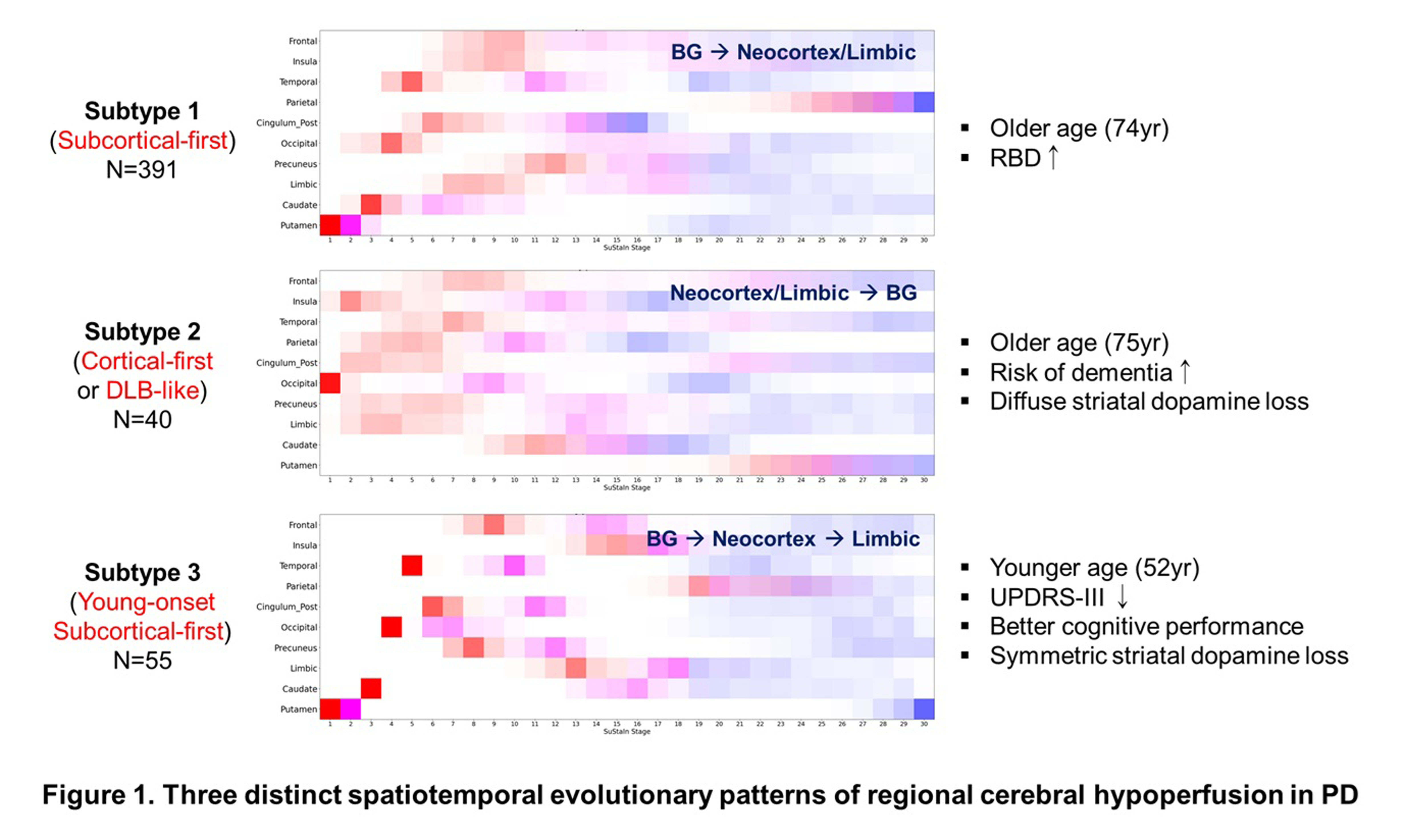
Figure 1
References: 1. Jeong, S.H., et al., Differential Implications of Cerebral Hypoperfusion and Hyperperfusion in Parkinson’s Disease. Mov Disord, 2023. 38(10): p. 1881-1890.
2. Chun, M.Y., et al., Hippocampal Perfusion Affects Motor and Cognitive Functions in Parkinson Disease: An Early Phase (18) F-FP-CIT Positron Emission Tomography Study. Ann Neurol, 2024. 95(2): p. 388-399.
3. Chun, M.Y., et al., Hypoperfusion in Alzheimer’s Disease-Prone Regions and Dementia Conversion in Parkinson’s Disease. Clin Nucl Med, 2024. 49(6): p. 521-528.
4. Peng, S., et al., Dynamic (18)F-FPCIT PET: Quantification of Parkinson’s disease metabolic networks and nigrostriatal dopaminergic dysfunction in a single imaging session. J Nucl Med, 2021. 62(12): p. 1775-82.
5. Young, A.L., et al., Uncovering the heterogeneity and temporal complexity of neurodegenerative diseases with Subtype and Stage Inference. Nat Commun, 2018. 9(1): p. 4273.
To cite this abstract in AMA style:
SJ. Chung, J. Lee, Y. Choi, J. Jang, HK. Na, PH. Lee. Uncovering the heterogeneity of spatiotemporal trajectories of neurodegeneration in Parkinson’s disease: an early-phase 18F-FP-CIT PET study [abstract]. Mov Disord. 2025; 40 (suppl 1). https://www.mdsabstracts.org/abstract/uncovering-the-heterogeneity-of-spatiotemporal-trajectories-of-neurodegeneration-in-parkinsons-disease-an-early-phase-18f-fp-cit-pet-study/. Accessed July 22, 2026.« Back to 2025 International Congress
MDS Abstracts - https://www.mdsabstracts.org/abstract/uncovering-the-heterogeneity-of-spatiotemporal-trajectories-of-neurodegeneration-in-parkinsons-disease-an-early-phase-18f-fp-cit-pet-study/