Session Information
Date: Monday, October 8, 2018
Session Title: Parkinson's Disease: Neuroimaging And Neurophysiology
Session Time: 1:15pm-2:45pm
Location: Hall 3FG
Objective: We aim to analyze cerebellar functional connectivity (FC) in PD patients with FOG (PD-FOG).
Background: Freezing of gait (FOG) is a disabling disorder frequently affecting patients with Parkinson’s disease (PD) and consists of transient inability to start or maintain locomotion [1]. The pathophysiological mechanism of FOG has not yet been identified. Although recent studies with magnetic resonance imaging (MRI) have shown alterations in several cortical and subcortical brain areas of patients with FOG, the cerebellum, a key structure in posture and gait control, has only been partially studied [2].
Methods: We recruited 15 PD-FOG, 16 PD without FOG (PD-nFOG) patients, and 16 age-matched healthy subjects (HS). The motor and cognitive impairment of patients was evaluated with validated clinical scales. FOG severity was assessed by the FOG Questionnaire (FOG-Q). Resting-state fMRI data were acquired using a high field magnet (3 Tesla, Siemens, Verio). By using the FSL software we located a seed region on the cerebellar locomotor region (CLR) and obtained FC maps of the CLR with the whole brain. The z scores of clusters showing significant differences in FC between PD-FOG patients and HS were correlated with scores at FOG-Q in SPSS toolbox.
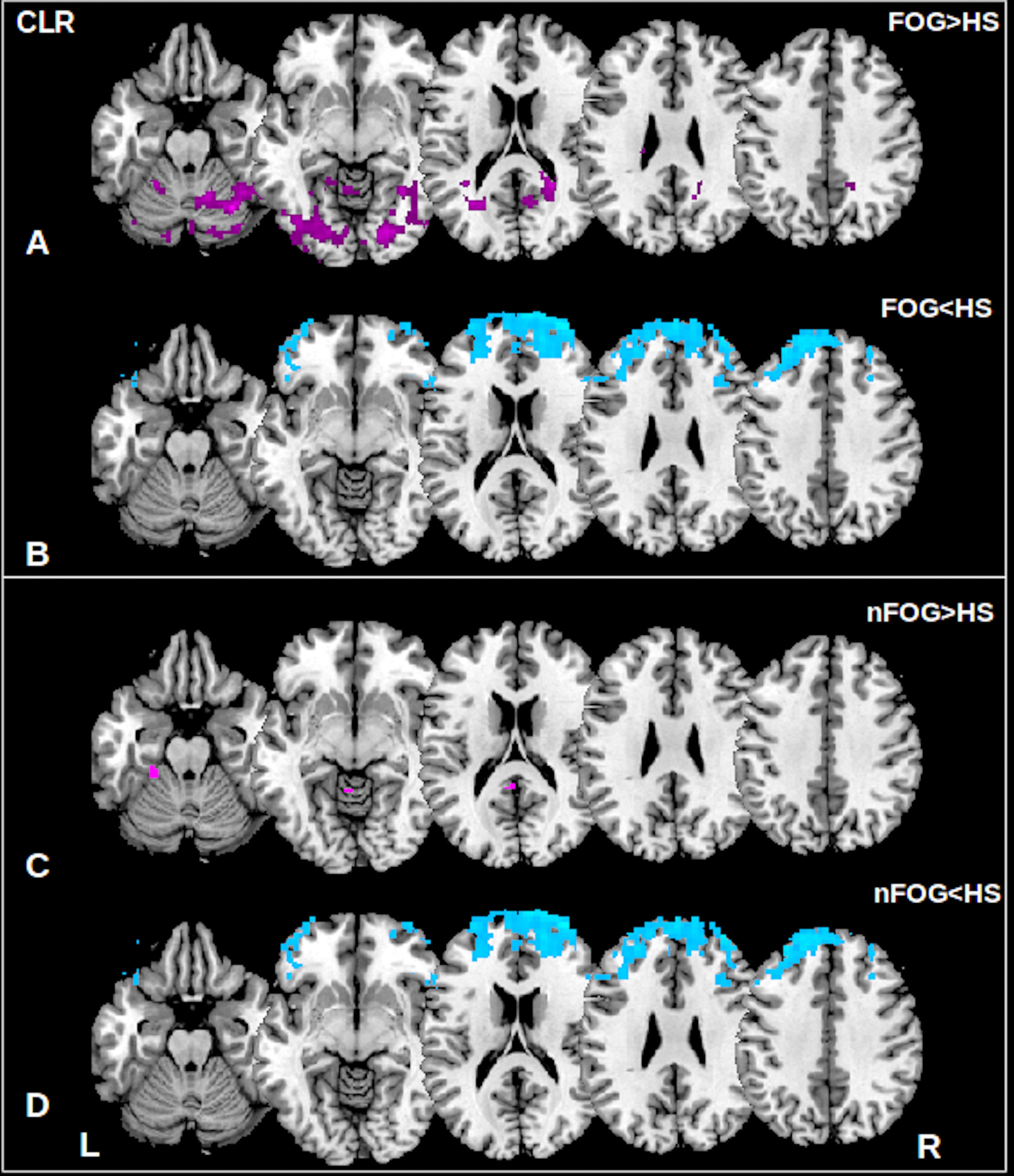
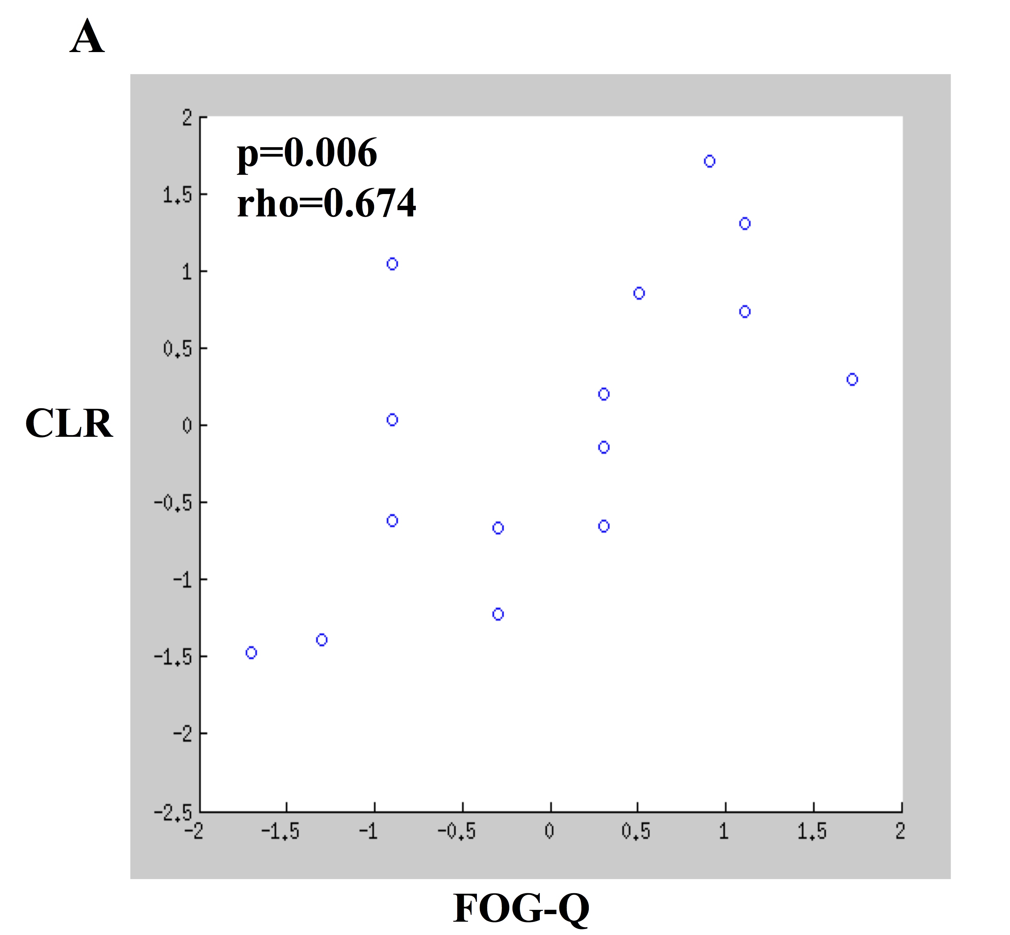
Results: The two subgroups of patients showed decreased FC of the CLR with the prefrontal cortex (superior, middle and inferior frontal gyrus) of both the cerebral hemispheres. In addition, only PD-FOG exhibited increased FC of CLR with the cerebellar vermis, cerebellar lobules left VI, IX, and right crus I-IV, VI, VII b as well as with the parieto-occipital cortex (both fusiform gyrus and lateral occipital cortex bilaterally and right lingual gyrus) [figure1]. FC of the CLR with cerebellar and posterior cortical areas in PD-FOG positively correlated with the FOG-Q score [figure2].
Conclusions: Our results suggest that the altered functional connectivity of the cerebellum may contribute to the pathophysiology of FOG; in particular, FC of the CLR, correlated with FOG severity, supporting the hypothesis that abnormal cerebellar function can contribute to the genesis of FOG in patients with PD.
References: 1. Giladi N, McDermott MP, Fahn S, Przedborski S, Jankovic J, Stern M, et al. Freezing of gait in PD: prospective assessment in the DATATOP cohort. Neurology. 2001;56:1712–21. 2. Tessitore A, Amboni M, Esposito F, Russo A, Picillo M, Marcuccio L, et al. Resting-state brain connectivity in patients with Parkinson’s disease and freezing of gait. Parkinsonism Relat Disord. 2012;18:781–7.
To cite this abstract in AMA style:
K. Bearti, A. Suppa, S. Pietracupa, N. Upadhyay, C. Giannì, G. Leodori, F. Di Biasio, N. Modugno, N. Petsas, G. Grillea, A. Zampogna, A. Berardelli, P. Pantano. Abnormal Functional Connectivity in Cerebellar Locomotor Region is associated with the severity of freezing of gait in patients with Parkinson’s disease [abstract]. Mov Disord. 2018; 33 (suppl 2). https://www.mdsabstracts.org/abstract/abnormal-functional-connectivity-in-cerebellar-locomotor-region-is-associated-with-the-severity-of-freezing-of-gait-in-patients-with-parkinsons-disease/. Accessed July 14, 2025.« Back to 2018 International Congress
MDS Abstracts - https://www.mdsabstracts.org/abstract/abnormal-functional-connectivity-in-cerebellar-locomotor-region-is-associated-with-the-severity-of-freezing-of-gait-in-patients-with-parkinsons-disease/


