Category: Parkinson's Disease: Neuroimaging
Objective: This study investigated whether brain-first and body-first Parkinson’s disease (PD) showed different pattern of subregional striatal dopamine loss and cerebral perfusion using dual-phase 18F-FP-CIT PET scans and their effect on cognitive outcome.
Background: A new hypothesis based on the initial site of pathology proposed two types in PD: brain-first versus body first PD. The difference in patterns of subregional striatal dopaminergic degeneration and cerebral perfusion between these two types has not been investigated yet.
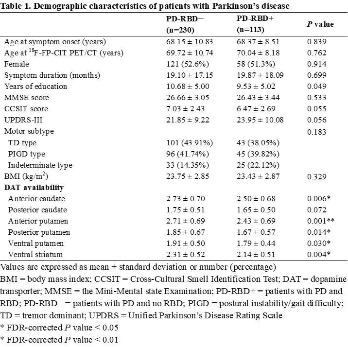
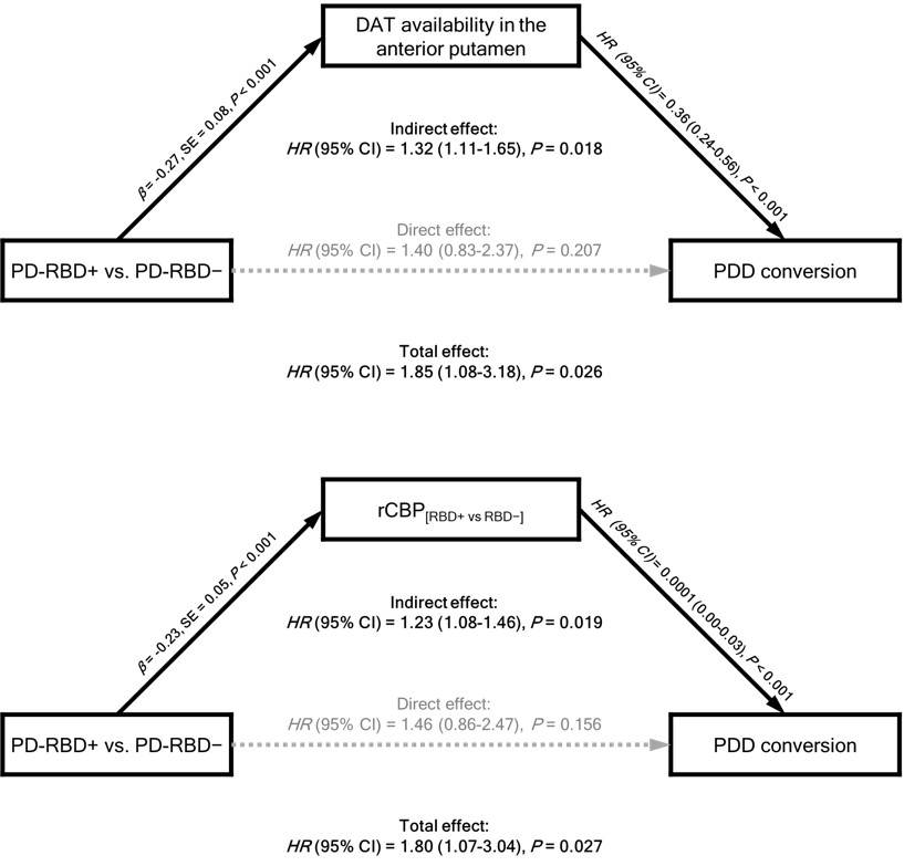
Method: We retrospectively reviewed data of 343 patients with de novo PD and 48 healthy controls from a tertiary hospital. We classified as PD with RBD (PD-RBD+) and without RBD (PD-RBD−) by using RBDSQ with a cutoff score of ≥6. Multivariable linear regression model was used to determine intergroup difference in the patterns of subregional dopamine transporter availability (DAT). We compared regional cerebral perfusion estimated using early-phase 18F-FP-CIT scans after a 1:1 propensity score matching. A Cox regression model was used to compare the risk of developing PD dementia (PDD) between the groups. A mediation analysis was used to explore whether the intergroup difference in imaging features mediate different cognitive outcome between the groups.
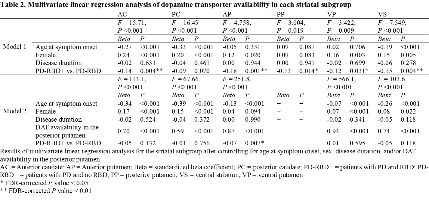
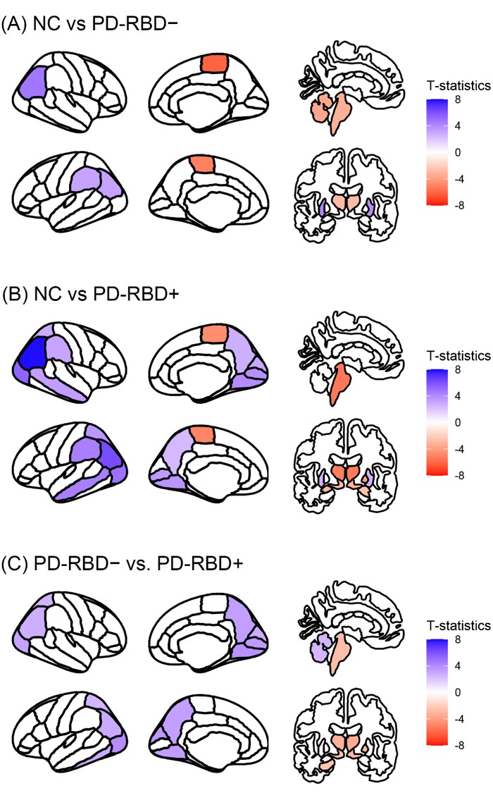
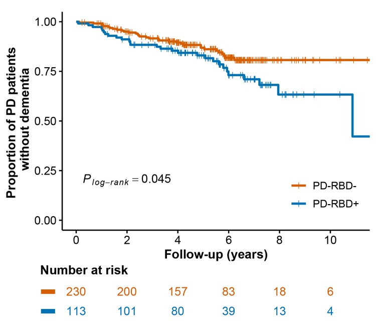
Results: After adjusting for covariates, DAT availability in the anterior putamen was significantly lower in the PD-RBD+ group than in the PD-RBD− group (Beta = -0.07, P = 0.007). In comparison of regional cerebral perfusion, regional perfusion in the bilateral parieto-occipital area, and left cerebellum were significantly lower in the PD-RBD+ group than those in the PD-RBD− group, while brainstem, left hippocampus, right pallidum, and bilateral thalamus and ventral diencephalon were vice versa. A survival analysis showed that the rate of PDD conversion was significantly higher in the PD-RBD+ group (hazard ratio = 1.78, 95% confidence interval = 1.07−2.99, P = 0.027) than the PD-RBD− group, which was fully mediated by DAT availability in the anterior putamen or parieto-occipital cerebral perfusion in mediation analyses.
Conclusion: This study demonstrated that the patterns of striatal subregional dopaminergic degeneration and regional cerebral perfusion differed between brain-first and body-first PD subtypes, and these differences fully mediate inter-subtype difference in cognitive outcome.
Table 1
Table 2
Table 3
Table 4
Figure 1
Figure 2
Figure 3
To cite this abstract in AMA style:
SH. Jeong, JS. Baik, PH. Lee, SJ. Chung. Association of Striatal Dopaminergic Depletion, Cerebral Perfusion, and Cognitive Outcome in Brain-first and Body-first Parkinson’s Disease [abstract]. Mov Disord. 2024; 39 (suppl 1). https://www.mdsabstracts.org/abstract/association-of-striatal-dopaminergic-depletion-cerebral-perfusion-and-cognitive-outcome-in-brain-first-and-body-first-parkinsons-disease/. Accessed July 18, 2025.« Back to 2024 International Congress
MDS Abstracts - https://www.mdsabstracts.org/abstract/association-of-striatal-dopaminergic-depletion-cerebral-perfusion-and-cognitive-outcome-in-brain-first-and-body-first-parkinsons-disease/

