Session Information
Date: Wednesday, June 7, 2017
Session Title: Neuroimaging (Non-PD)
Session Time: 1:15pm-2:45pm
Location: Exhibit Hall C
Objective: To investigate whether cerebral iron deposits in Wilson disease (WD) patients are associated with decreased serum ceruloplasmin oxidase activity and/or clinical severity.
Background: WD is a genetic disorder caused by mutation of ATP7B leading to systemic copper accumulation. Abnormal deposition of paramagnetic compounds in the deep gray matter (DGM) in WD patients were reported in previous MRI studies[1]. A post-mortem MRI histopathology correlation study in WD showed that hypointense signal on T2* weighted MRI in the DGM was correlated with iron concentration and with the number of iron-loaded macrophages, whereas no correlation was observed for the copper concentration[2]. Pathophysiological underpinnings and clinical implications of iron deposits in WD are unknown.
Methods: 26 patients with genetically confirmed neurologic WD on anti-copper treatment (16f/10m, 28-63 yrs, disease duration 1-46 yrs) and 16 healthy controls (9f/7m, 28-60 yrs) were investigated. Clinical severity was assessed using the Unified Wilson Disease Rating Scale (UWDRS). Serum ceruloplasmin oxidase activity was measured using the o-dianysine assay. MRI was performed on a 3T scanner using a 12‑channel head coil. Reconstruction of quantitative susceptibility maps (QSM) from 3D multi gradient recalled echo sequence (TR=40 ms; six TEs 5.22-34.64 ms; FA=15°; voxel resolution = 0.8×0.8×2 mm2) consisted of continuous Laplacian unwrapping, spherical mean value property based background field removal, and non-linear MEDIN inversion. Mean QSM values, considered as a measure of tissue iron concentration, were calculated in: globus pallidus (GP), Putamen (Put), Caudate nucleus (CN), and Thalamus (Th). Mann-Whitney U test was used for group comparisons, correlations were examined by Spearman coefficient.
Results: In WD patients, significantly higher susceptibility values were detected in all DGM structures [table1]. Mean neurological UWDRS subscore was 22.5 (1-56); it did not correlate with susceptibility values in any brain structure. Although all WD patients had low ceruloplasmin levels, susceptibility values did not correlate with the ceruloplasmin oxidase activity [table2].
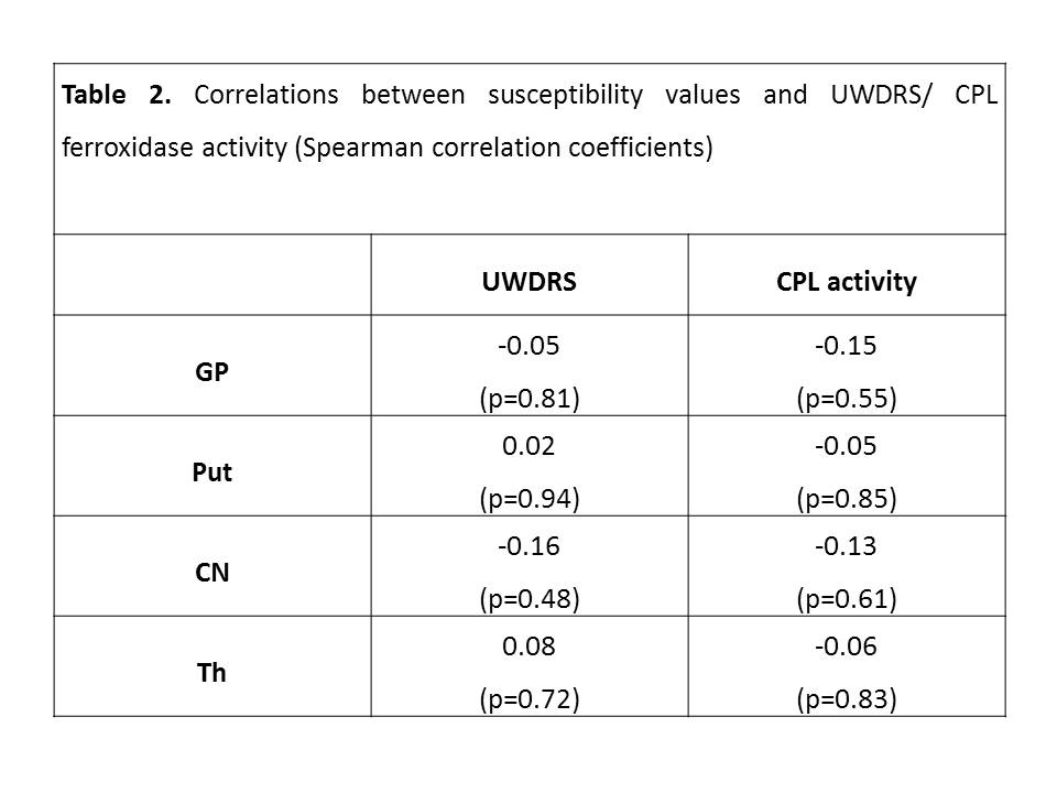
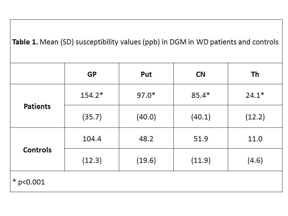
Conclusions: Increased susceptibility values in DGM indicate that WD patients have diffusely increased cerebral iron levels. These iron deposits appear to be not causally related to decreased ferroxidase activity nor associated with clinical severity.
References: 1. Fritzsch D et al., Invest Radiol 2014;49(5):299-306.
2. Dusek P et al. Neuropathol Appl Neurobiol 2016; doi: 10.1111/nan.12341.
To cite this abstract in AMA style:
P. Dusek, M. Dezortova, V. Herynek, J. Acosta-Cabronero, L. Kotackova, D. Zahorakova, S. Robinson, F. Jiru, R. Bruha, Z. Marecek, M. Hajek. Brain iron accumulation in Wilson disease measured by quantitative susceptibility mapping [abstract]. Mov Disord. 2017; 32 (suppl 2). https://www.mdsabstracts.org/abstract/brain-iron-accumulation-in-wilson-disease-measured-by-quantitative-susceptibility-mapping/. Accessed March 19, 2026.« Back to 2017 International Congress
MDS Abstracts - https://www.mdsabstracts.org/abstract/brain-iron-accumulation-in-wilson-disease-measured-by-quantitative-susceptibility-mapping/