Category: Parkinson's Disease: Neuroimaging
Objective: To detect patients with Parkinson’s disease that have REM sleep behavior disorder (RBD) and perform brain MRI volumetry analysis.
Background: Parkinson’s disease is classically related to loss of dopaminergic neurons due to degeneration of substantia nigra. It has been reported that patients with Parkinson’s disease (pwPD) also have reduced volume of gray matter in frontal lobes [1], hipoccampus, cingulate gyrus, upper temporal gyrus, caudate nucleus, putamen [2]. Sleep – wake disorders are among the main non-motor symptoms (NMS) that occur for the patients with Parkinson’s disease (pwPD), affecting 60-90% of them [3]. Using brain MRI volumetry analysis it has been detected that patients suffering from REM sleep behavior disorder (RBD) may have reduced volume of reticular formation, hypothalamus, thalamus, putamen, amygdala, frontal part of cingulate gyrus [4]. Recent studies show that RBD occur 10-15 years before PD specific motor symptoms evolve and is associated with increased risk of developing other neurodegenerative diseases [5].
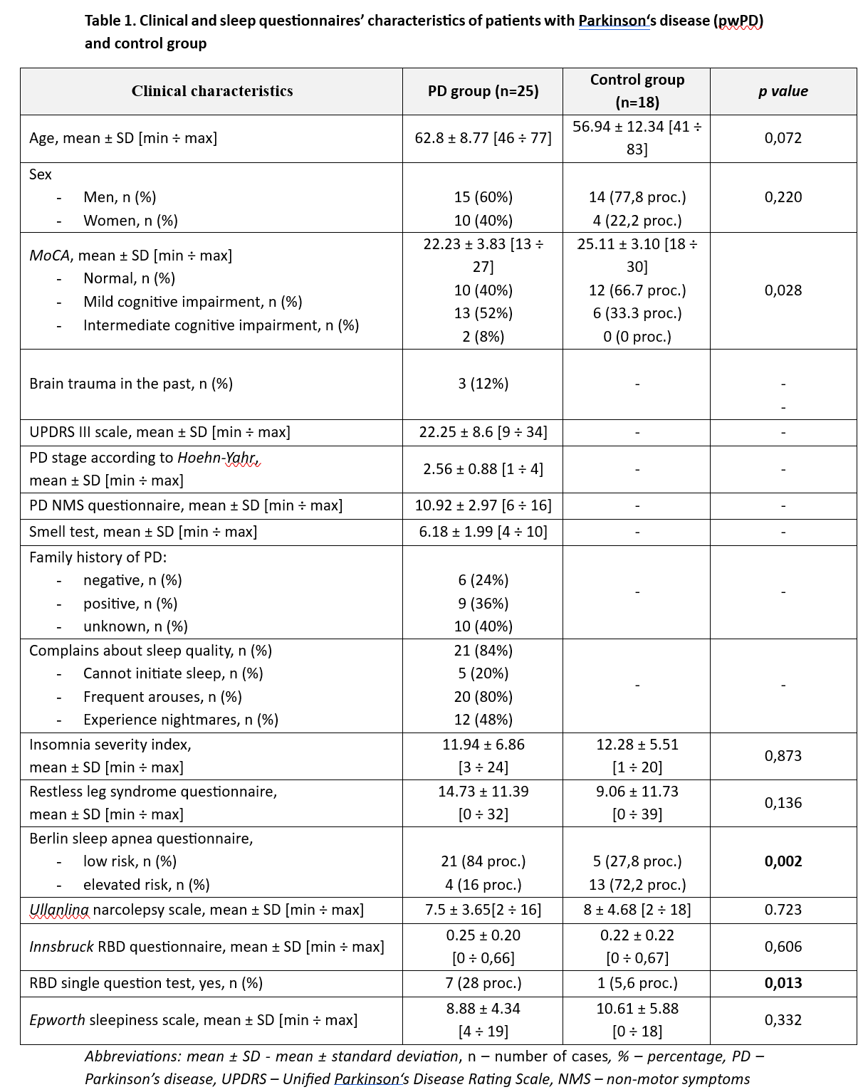
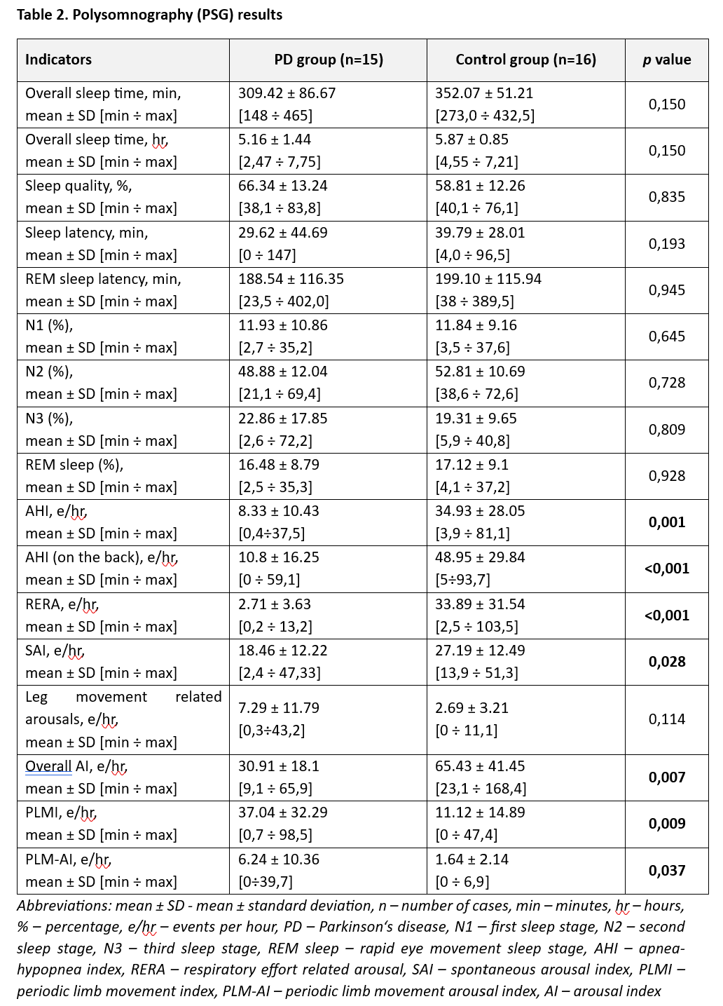
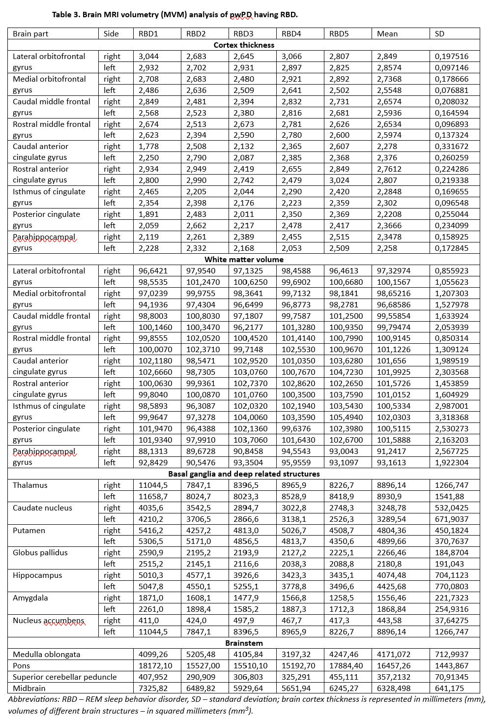
Method: We involved 43 patients (25 patients with PD and 18 control group patients who underwent clinical evaluation of motor (UPDRS III scale) and non-motor symptoms (NMS) (Table 1): NMS-quest, Sniffin’Sticks-12 (SS-12) test, Montreal cognitive assessment (MoCA), sleep questionnaires. 15 PD and 16 control group patients underwent polysomnography (PSG) and were evaluated for objective sleep disorders (Table 2). After PSG, patients confirmed having RBD were evaluated using brain MRI volumetry analysis (n=5). Automated voxel-based morphometry of brain cortex and subcortical structures’ analysis was performed using open source FreeSurfer program.
Results: Majority of our PD cohort got the PSG-based diagnosis of RBD (75%). In addition to sleep-related symptoms, they were more frequently suffering from other NMS. The study is ongoing and further controlled brain volumetry data will be added (Table 3).
Conclusion: It is a pilot study and only pwPD having RBD were analyzed. Brain MRI volumetry analysis is an exciting tool for evaluating various brain areas and provides deeper understanding about the nature of PD.
References: 1. Burton EJ, McKeith IG, Burn DJ, Williams ED, O’Brien JT. Cerebral atrophy in Parkinson’s disease with and without dementia: a com-parison with Alzheimer’s disease, dementia with Lewy bodies and controls. Brain. 2004;127:791–800.
2. Summerfield C, Junqué C, Tolosa E, Salgado-Pineda P, Gómez-Ansón B, Martí MJ, et al. Structural Brain Changes in Parkinson Disease With Dementia. Arch Neurol. 2005; 62:281.
3. Chahine, L. M., Amara, A. W., and Videnovic, A. (2017). A systematic review of the literature on disorders of sleep and wakefulness in Parkinson’s disease from 2005 to 2015. Sleep Med Rev. 35, 33–50.
4. Boucetta S, Salimi A, Dadar M, Jones BE, Collins DL, Dang-Vu TT. Structural Brain Alterations Associated with Rapid Eye Movement Sleep Behavior Disorder in Parkinson’s Disease. Sci Rep. 2016;6: 26782.
5. Barber TR, Lawton M, Rolinski M, et al. Prodromal parkinsonism and neurodegenerative risk stratification in REM sleep behavior disorder. Sleep 2017;40(8):zsx071.
To cite this abstract in AMA style:
T. Vanagas, G. Paulekiene, A. Radziunas, R. Balnyte, E. Pajediene, E. Paulekas. Brain MRI volumetry analysis for patients with Parkinson’s disease suffering from REM sleep behavior disorder: a pilot study [abstract]. Mov Disord. 2023; 38 (suppl 1). https://www.mdsabstracts.org/abstract/brain-mri-volumetry-analysis-for-patients-with-parkinsons-disease-suffering-from-rem-sleep-behavior-disorder-a-pilot-study/. Accessed June 14, 2026.« Back to 2023 International Congress
MDS Abstracts - https://www.mdsabstracts.org/abstract/brain-mri-volumetry-analysis-for-patients-with-parkinsons-disease-suffering-from-rem-sleep-behavior-disorder-a-pilot-study/