Category: Ataxia
Objective: The aim of our work is to create a novel protocol for manual subsegmentation of the human brainstem for creating accurate brainstem labels for volumetric analysis. In utilizing anatomical landmarks for subdividing the brainstem into subregions we aim to receive reproduceable results, allowing improved comparability of data. We applied the protocol on 30 subjects – 10 healthy controls, 10 pre-symptomatic SCA3 patients (preSCA3) and 10 symptomatic SCA3 patients (SCA3).
Background: SCA3 is an inherited neurodegenerative disease which leads to atrophy of brain regions mainly associated with coordination of movements. Atrophy of these regions already occurs before onset of symptoms. Assessment of atrophied regions could present as an important biomarker. Biomarkers are important for understanding disease progression in SCA3. Manual Subsegmentation is a method of subdividing specific organ regions in imaging to allow for several applications like volumetric analysis. It allows accurate delineation of brain structures and remains the gold standard in comparison to fully automated approaches. Currently there are tools for fully automated subsegmentation, especially for supratentorial regions. However only limited options for brainstem subsegmentation exist, often not including all of the cerebellar peduncles. Therefore, we propose a modified protocol that can serve as the basis for gathering new ground truth data for model training.
Method: T1-weighted 3T-MRI-images with an isotropic voxel-size of 0.8mm were used. Images were manually segmented by a trained physician using ITK-Snap. Following a modified subsegmentation manual based on robust anatomical landmarks to minimize subjective rating. The brainstem was subdivided into five labels: Midbrain, Pons, Medulla Oblongata, SCP and the merged label of MCP and ICP.
Results: Our results show differences in volume between healthy controls, preSCA3 and SCA3 patients. With the exception of the SCP, all of the five regions of interest show a difference in volume among all groups, with SCA3 patients having the lowest volumes and preSCA3 patients having the second lowest. The most striking volume difference occurred in pons.
Conclusion: Manual subsegmentation of brainstem regions is a valuable tool and provides promising biomarkers for assessment of disease progression in SCA3 as shown in this study. We were able to show differences in volumes among all groups, which reflects the reality of disease.
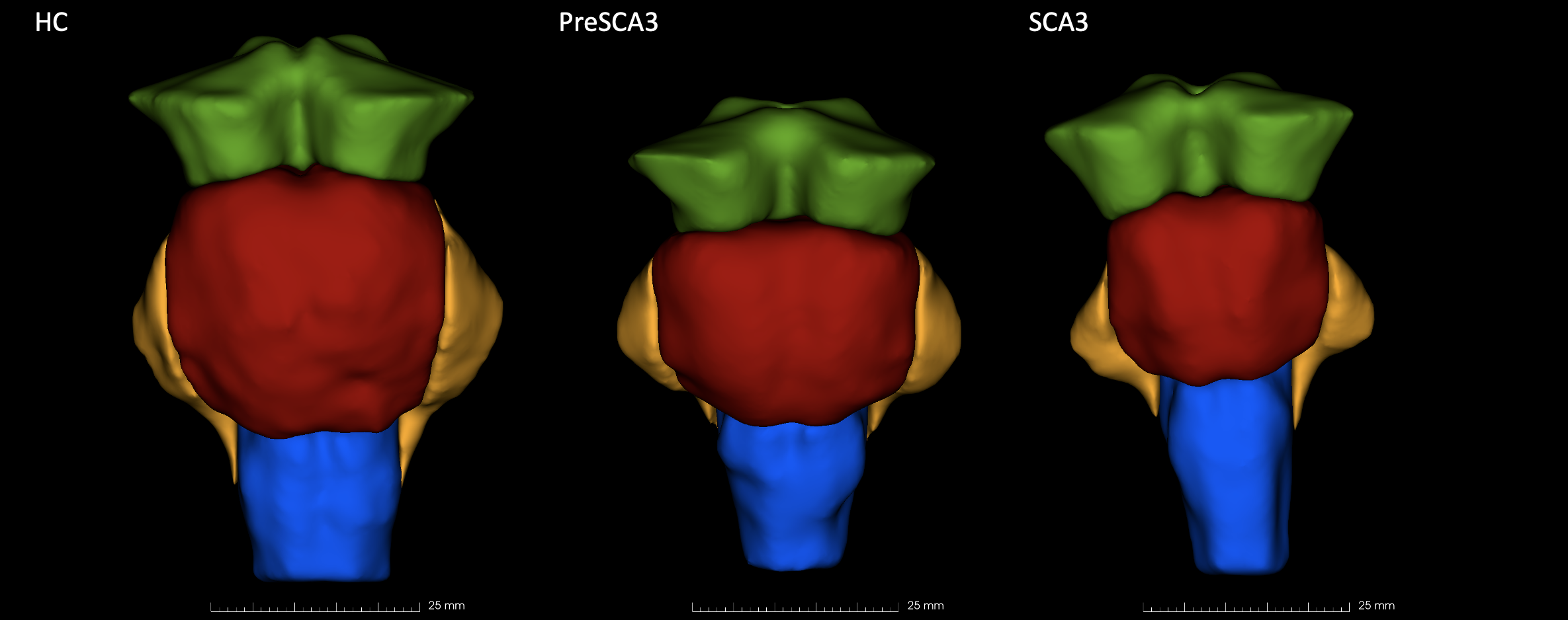
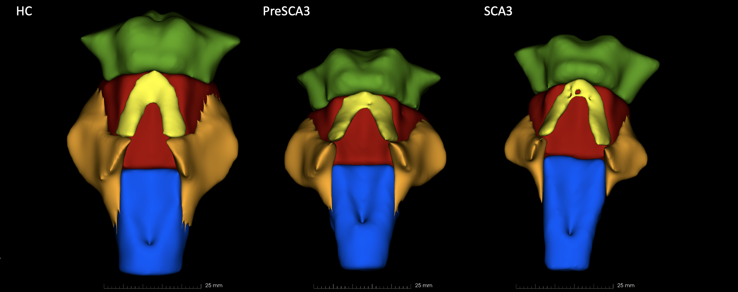
Anterior view comparison in 3D Slicer
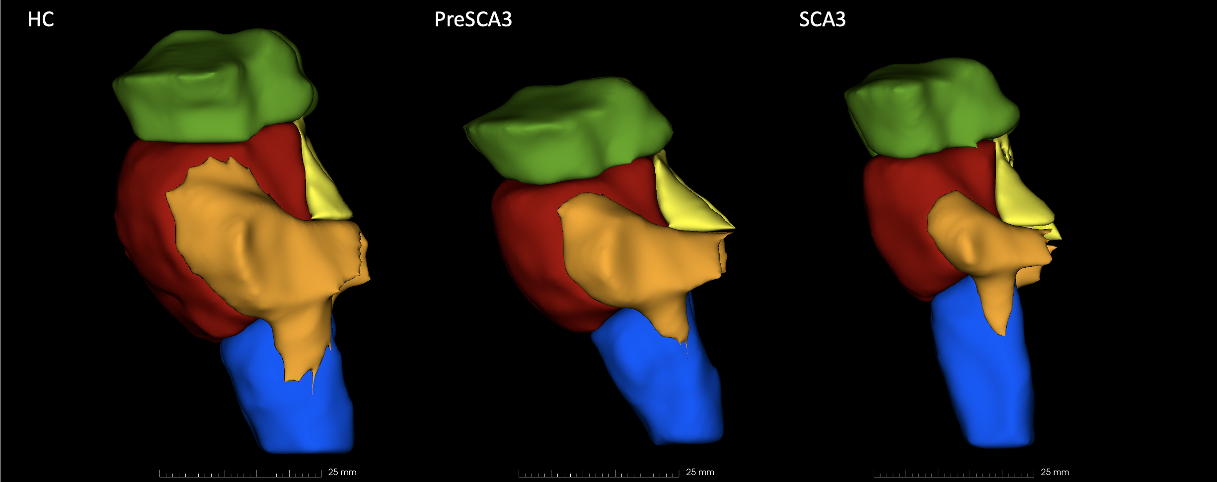
Lateral view comparison in 3D Slicer
Posterior view comparison in 3D Slicer
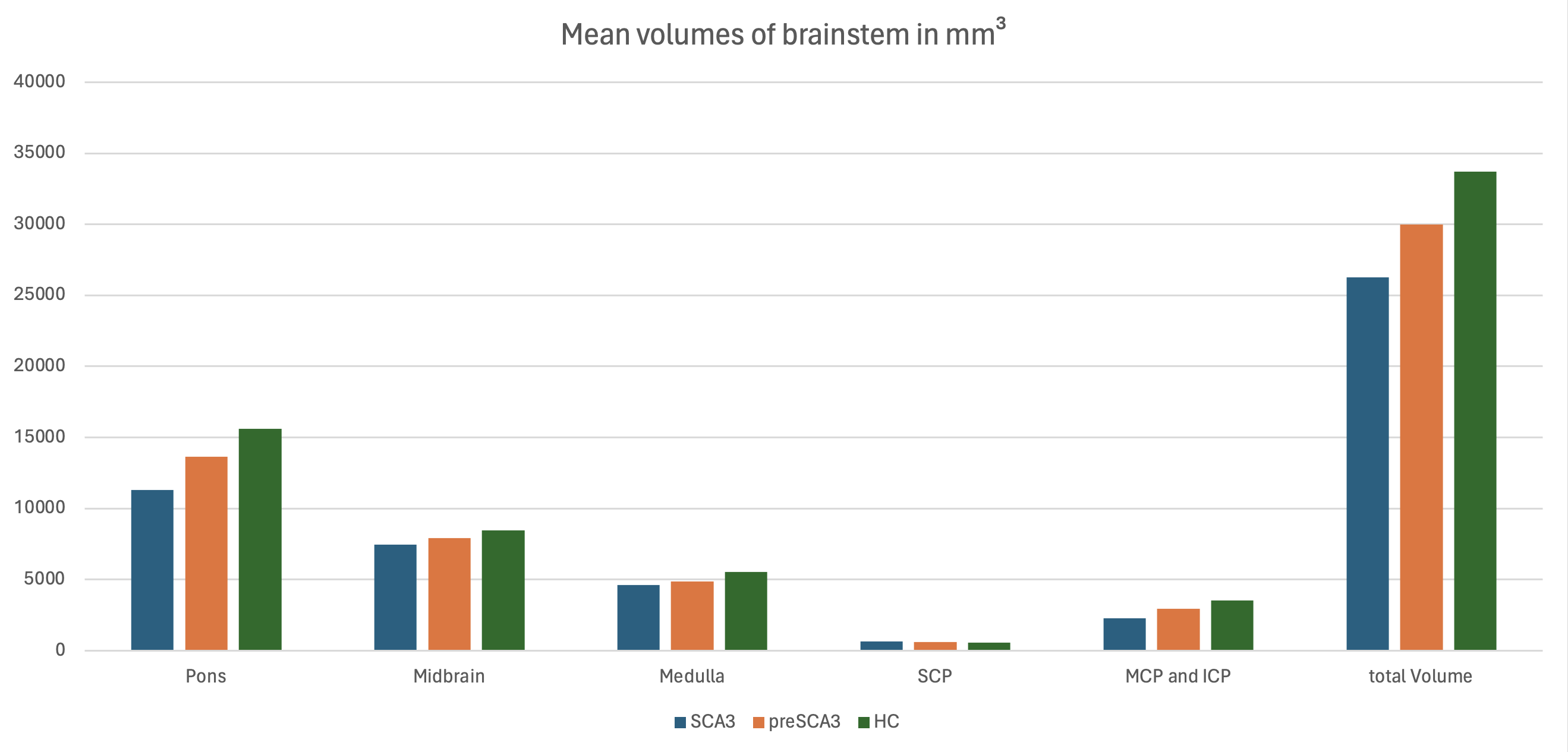
Volume comparison in mm³
References: Klockgether T, Mariotti C, Paulson HL. Spinocerebellar ataxia. Nat Rev Dis Primers. 2019 Apr 11;5(1):24. doi: 10.1038/s41572-019-0074-3. PMID: 30975995.
Henschel L, Conjeti S, Estrada S, Diers K, Fischl B, Reuter M, FastSurfer – A fast and accurate deep learning based neuroimaging pipeline, NeuroImage 219 (2020), 117012. https://doi.org/10.1016/j.neuroimage.2020.117012
Faber J, Kügler D, Bahrami E, Heinz LS, Timmann D, Ernst TM, Deike-Hofmann K, Klockgether T, van de Warrenburg B, van Gaalen J, Reetz K, Romanzetti S, Oz G, Joers JM, Diedrichsen J; ESMI MRI Study Group; Reuter M. CerebNet: A fast and reliable deep-learning pipeline for detailed cerebellum sub-segmentation. Neuroimage. 2022 Dec 1;264:119703. doi: 10.1016/j.neuroimage.2022.119703. Epub 2022 Oct 27. PMID: 36349595; PMCID: PMC9771831.
Iglesias JE, Van Leemput K, Bhatt P, Casillas C, Dutt S, Schuff N, Truran-Sacrey D, Boxer A, Fischl B; Alzheimer’s Disease Neuroimaging Initiative. Bayesian segmentation of brainstem structures in MRI. Neuroimage. 2015 Jun;113:184-95. doi: 10.1016/j.neuroimage.2015.02.065. Epub 2015 Mar 14. PMID: 25776214; PMCID: PMC4434226.
Weier K, Fonov V, Lavoie K, Doyon J, Collins DL. Rapid automatic segmentation of the human cerebellum and its lobules (RASCAL)–implementation and application of the patch-based label-fusion technique with a template library to segment the human cerebellum. Hum Brain Mapp. 2014 Oct;35(10):5026-39. doi: 10.1002/hbm.22529. Epub 2014 Apr 28. PMID: 24777876; PMCID: PMC6869487.
Paul A. Yushkevich, Joseph Piven, Heather Cody Hazlett, Rachel Gimpel Smith, Sean Ho, James C. Gee, and Guido Gerig. User-guided 3D active contour segmentation of anatomical structures: Significantly improved efficiency and reliability. Neuroimage 2006 Jul 1;31(3):1116-28.
Kikinis R, Pieper SD, Vosburgh K (2014) 3D Slicer: a platform for subject-specific image analysis, visualization, and clinical support. Intraoperative Imaging Image-Guided Therapy, Ferenc A. Jolesz, Editor 3(19):277–289 ISBN: 978-1-4614-7656-6 (Print) 978-1-4614-7657-3 (Online)
To cite this abstract in AMA style:
K. Teichmann. Brainstem Subsegmentation in Spinocerebellar Ataxia Type 3 (SCA3) [abstract]. Mov Disord. 2025; 40 (suppl 1). https://www.mdsabstracts.org/abstract/brainstem-subsegmentation-in-spinocerebellar-ataxia-type-3-sca3/. Accessed April 7, 2026.« Back to 2025 International Congress
MDS Abstracts - https://www.mdsabstracts.org/abstract/brainstem-subsegmentation-in-spinocerebellar-ataxia-type-3-sca3/