Category: Parkinson's Disease: Neuroimaging
Objective: To investigate (1)longitudinal change and (2)cross-sectional vendor differences in neuromelanin MRI in substantia nigra pars compacta(SNpc) in PD patients.
Background: Neuromelanin(NM) MRI is a proposed biomarker of dopaminergic neurodegeneration in the SNpc[1]. We performed post-hoc analyses on data acquired from a large multi-center clinical trial (SPARK:NCT03318523) to characterize NM-MRI sensitivity to longitudinal change and cross-vendor variability.
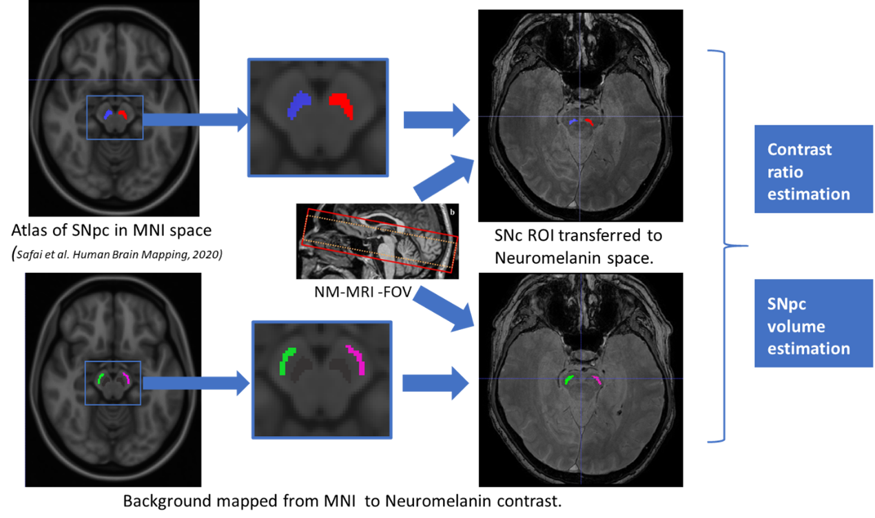
Method: 112 subjects from the SPARK study with PD diagnosis ≤3 yrs, a modified H&Y score ≤ 2.5, no symptomatic treatment within 12 weeks, and confirmed abnormal striatal DaTscan uptake were included. Subjects were followed longitudinally with 3T MRI acquired at screening, 24,52 and 96 weeks. NM-MRI was acquired using a 3DGRE sequence to cover the mid-brain with TR/TE=55/~6.5ms, 0.62 in-slice resolution, 1.3mm slice thickness. 3D-T1-weighted scans were acquired at 1mm3 resolution. The following distribution of subjects had scans that met quality control criteria for all time-points: manufacturer1(18 subjects), manufacturer2(12 subjects), and manufacturer3(82 subjects). The SNpc atlas from [2] was mapped to the NM space using ANTS [3] between MNI and the subject’s T1 followed by an affine registration between T1 and NM. SNpc volume and contrast ratio(CR) were then computed. The reference region(RR) [figure1] was created by editing the motor region from [4] mapped to NM-MRI in a similar fashion. CRs ((ISN-IRR)/IRR) were computed from the mean ROI intensity(I). The change in volume and CR at week 24, 52, and 96 was computed for each subject. An ANOVA model was employed to test for significant volume and CR change. To evaluate cross-sectional vendor differences, a MANCOVA model was used with manufacturer as the variable of interest and age and gender as covariates.
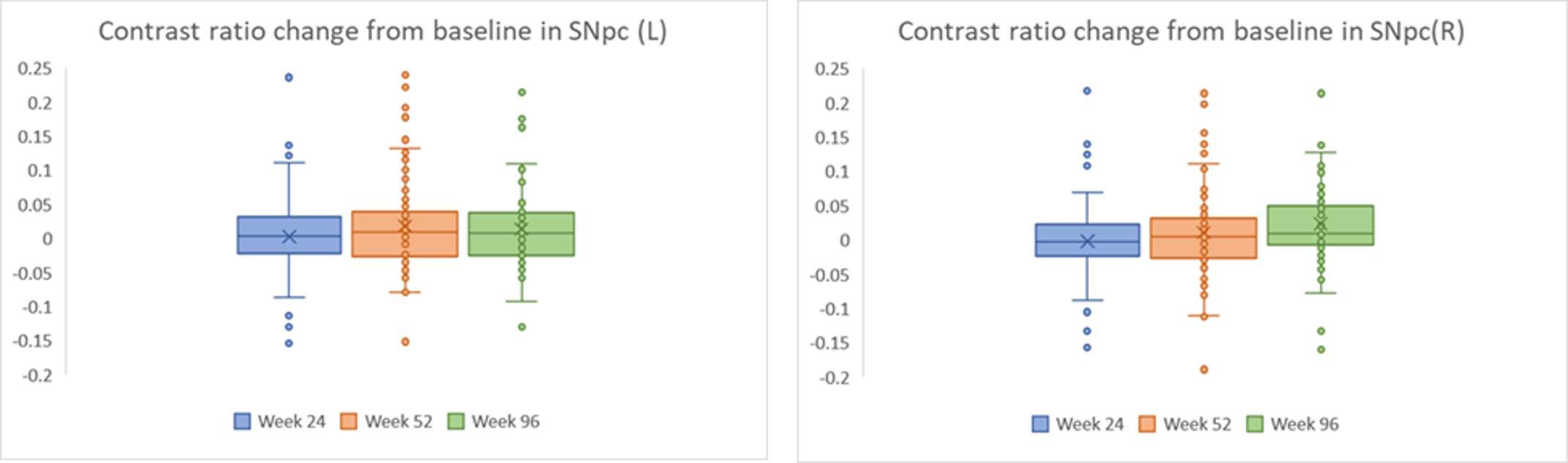
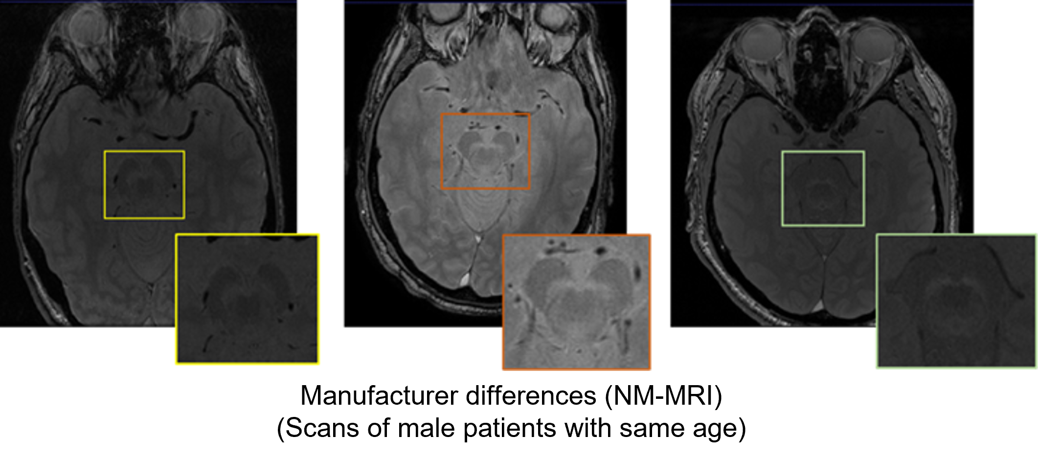
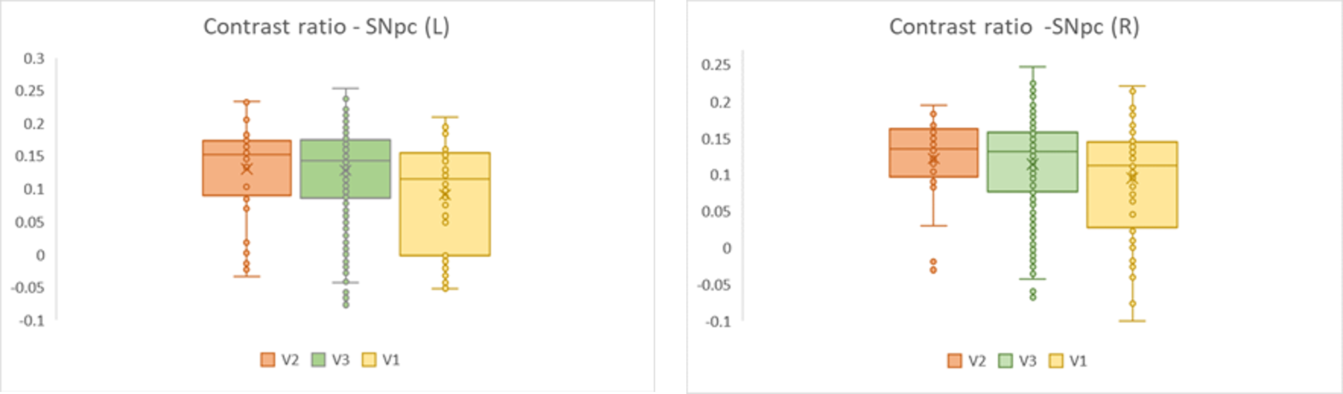
Results: Overall, CR significantly reduced on left side at week 52 from baseline, and bilaterally at week 96 [figure 2][table1]. SNpc volumes did not show any significant change. Manufacturer2 and 3 yielded significantly higher CR than manufacturer1, however manufacturer3 demostrated high variability[figure3][figure4][table2].
Conclusion: Longitudinal effects were generally small and variable, with inter-vendor differences. While left SNpc compared to right showed greater longitudinal change, additional work will be needed to establish NM-MRI as a viable marker of PD progression.


References: 1. Gaurav, R., Yahia‐Cherif, L., Pyatigorskaya, N., Mangone, G., Biondetti, E., Valabrègue, R., … & Lehéricy, S. (2021). Longitudinal changes in neuromelanin MRI signal in Parkinson’s disease: A progression marker. Movement Disorders, 36(7), 1592-1602.
2. Safai, A., Prasad, S., Chougule, T., Saini, J., Pal, P. K., & Ingalhalikar, M. (2020). Microstructural abnormalities of substantia nigra in Parkinson’s disease: a neuromelanin sensitive MRI atlas-based study. Human brain mapping, 41(5), 1323-1333.
3. Avants, Brian B., Nick Tustison, and Gang Song. “Advanced normalization tools (ANTS).” Insight j 2.365 (2009): 1-35.
4. Hua, K., Zhang, J., Wakana, S., Jiang, H., Li, X., Reich, D. S., … & Mori, S. (2008). Tract probability maps in stereotaxic spaces: analyses of white matter anatomy and tract-specific quantification. Neuroimage, 39(1), 336-347.
To cite this abstract in AMA style:
M. Ingalhalikar, H. Pham, L. Bracoud, RM. Hutchison, K. Evans, T. Dam, M. Sampat, J. Schaerer, C. Conklin, J. Suhy, D. Scott. Characterization of neuromelanin MRI as a potential Parkinson’s Disease biomarker in a multi-site longitudinal study [abstract]. Mov Disord. 2022; 37 (suppl 2). https://www.mdsabstracts.org/abstract/characterization-of-neuromelanin-mri-as-a-potential-parkinsons-disease-biomarker-in-a-multi-site-longitudinal-study/. Accessed May 6, 2026.« Back to 2022 International Congress
MDS Abstracts - https://www.mdsabstracts.org/abstract/characterization-of-neuromelanin-mri-as-a-potential-parkinsons-disease-biomarker-in-a-multi-site-longitudinal-study/