Session Information
Date: Tuesday, June 21, 2016
Session Title: Parkinson's disease: Pathophysiology
Session Time: 12:30pm-2:00pm
Location: Exhibit Hall located in Hall B, Level 2
Objective: In this study, we aimed to investigate the neural underpinning of freezing of gait (FOG) in Parkinson’s disease (PD), especially the role of the cerebral cortex in development of FOG.
Background: FOG is an episodic inability to generate effective stepping, and one of the most disabling features of patients with PD. Although previous findings suggests that the supraspinal gait-related neural network may contribute the FOG in PD, the primary lesion site is still unclear.
Methods: We recruited 23 PD patients with freezing and age-matched 13 healthy subjects. After we evaluated clinical status, all participants were evaluated their anatomical and functional network status using the diffusion tensor MR imaging and resting state functional MRI (rsfMRI) with 3T-MRI scanner (GE medical systems, Milwaukee, WI). First, we evaluated the voxel-by-voxel regression analysis to determine the brain area where fractional anisotropy (FA) value showed significant correlation with freezing severity. After determined the significant cluster in the brainstem tegmentum, we compared the functional connectivity status from the tegmentum area using the significant clusters as seed. We also estimated the cortical area where shows the significant correlation between the freezing severity and the functional connectivity status from the tegmentum area.
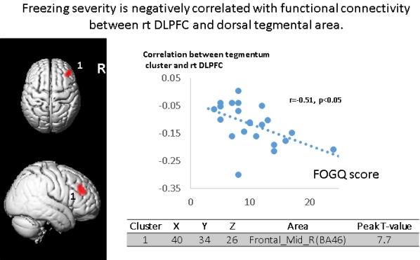
Results: Our findings revealed that the dorsal tegmentum area in the midbrain showed the significant correlation between freezing severity and FA values. The functional connectivity analysis revealed that several cortical regions including the bilateral intraparietal sulcus, right prefrontal cortex, bilateral cerebellar hemisphere, right temporal cortex, right thalamus, and right medial sensorimotor cortex showed reduced functional connectivity with the dorsal tegmental area in PD. Regression analysis showed the significant negative correlation between freezing severity and the functional connectivity between right dorsolateral prefrontal cortex and tegmentum area.
Conclusions: Our findings suggested that the dorsal tegmental area would be an possible “key structure” for freezing of gait and the right dorsolateral prefrontal cortex might compensate the deteriorated tegmental function.
To cite this abstract in AMA style:
M. Mihara, H. Otomune, H. Fujimoto, K. Konaka, Y. Watanabe, H. Mochizuki. Cortical role in the freezing of gait in Parkinson’s disease [abstract]. Mov Disord. 2016; 31 (suppl 2). https://www.mdsabstracts.org/abstract/cortical-role-in-the-freezing-of-gait-in-parkinsons-disease/. Accessed July 15, 2025.« Back to 2016 International Congress
MDS Abstracts - https://www.mdsabstracts.org/abstract/cortical-role-in-the-freezing-of-gait-in-parkinsons-disease/
