Category: Parkinson's Disease: Neuroimaging
Objective: To explore the feasible of depicting the nigrostriatal pathway (NSP), dentate rubro thalamic (DRTT) and hyperdirect pathway (HDP) for the postoperative PD patients with DBS on or off.
Background: Deep brain stimulation (DBS) has become an effective therapy for Parkinson’s disease (PD). It is of great value to depict the white matter tracts after DBS [1]. Preliminary evidence supports the safety of diffusion weighted tractography in postoperative patients, but it has not been reported whether the different states of DBS (on/off) affect the fiber bundle reconstruction, and whether it is feasible to depict the NSP, DRTT and HDP, which involved with control of motor behavior or be responsible in the pathophysiology of tremor in PD patients[2-4].
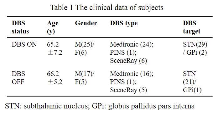
Method: A total of 31 postoperative PD cases were collected on a 1.5 T Siemens Aera scanner with a 16-channel head coil (Table 1). All the 31 patients were scanned with the DBS device on and 22 of them were rescanned with DBS off using the same DTI protocol: b value=1000 s/mm2, dir=64, TR=6500ms, TE=126ms, voxel size=2×2×2mm3, SAR= 0.34±0.02 w/kg, lower than the recommended threshold [5]. Probabilistic tractography of the NSP, HDP, and DRTT were performed using Probtrackx 2.0 [6-8]. Statistical analyses were conducted in SPSS 22.0. Findings with a p value of < 0.05 were considered significant.
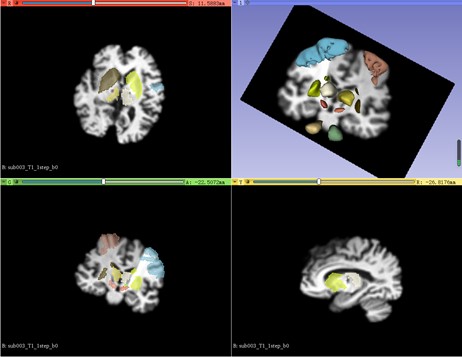
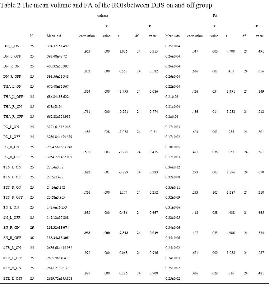
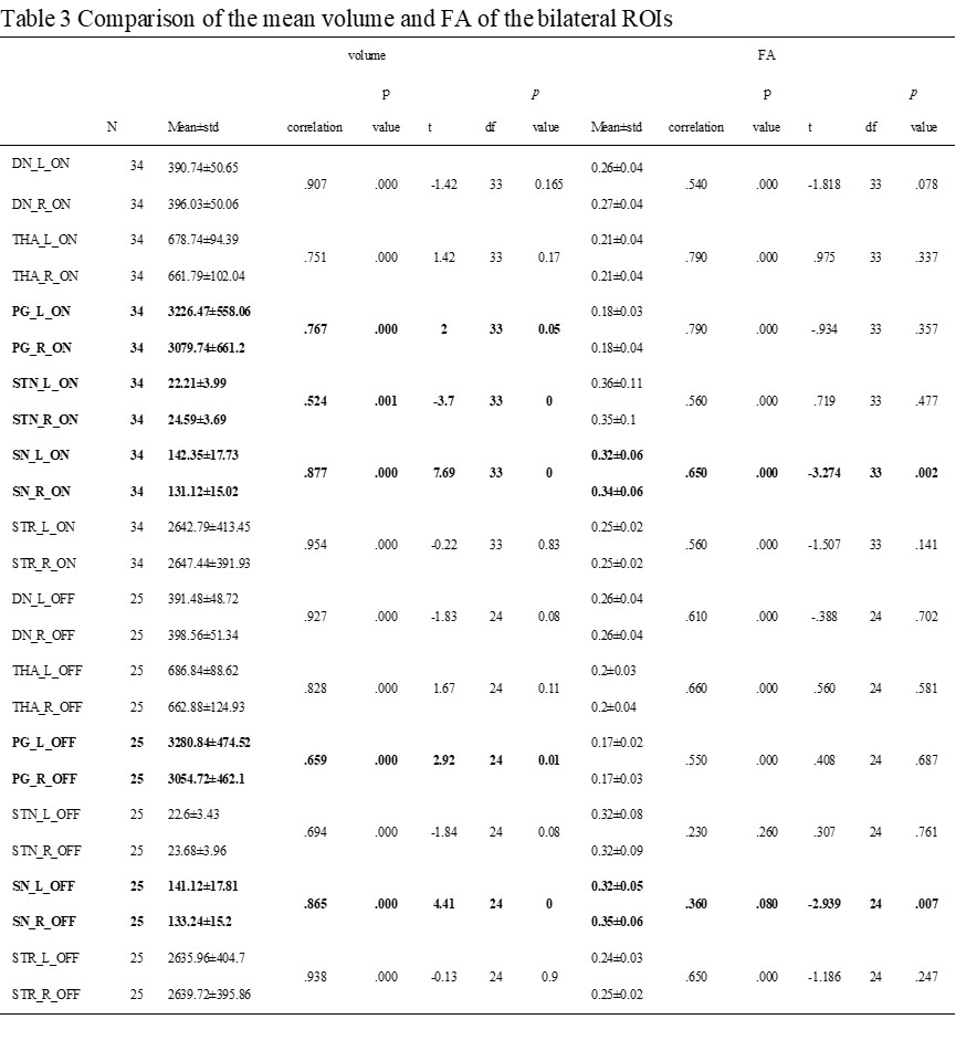
Results: A representative case and segmented ROIs were shown in the Figure 1. The mean values of volumes and FA of the ROIs were displayed in Table 2 and 3. The volumes of the ROIs were not differed significantly between the DBS on and off groups except for right substantia nigra.
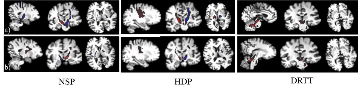
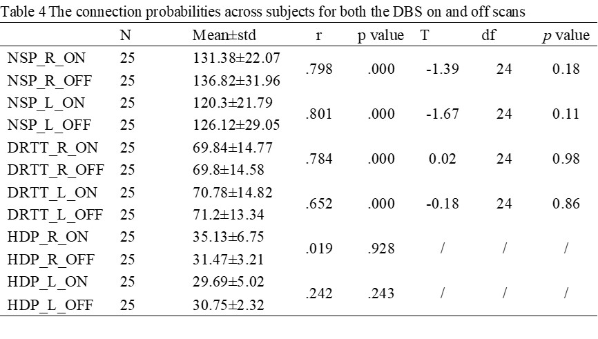
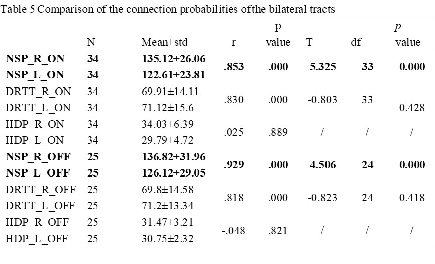
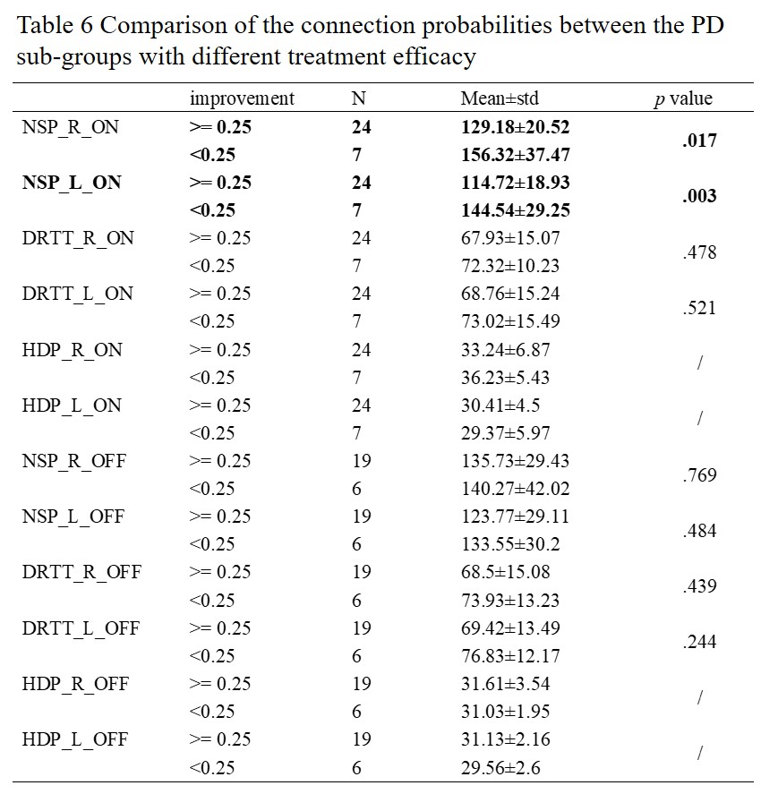
Probabilistic fiber tracking detected the NSP, DRTT and HDP in all hemispheres, as shown in Figure 2. It appears that structures not involving the cortex had the highest retention of tracts in postoperative imaging, with the NSP being the best retained, followed by the DRTT and the HDP. The connection probability average across subjects for both the DBS on and off scans could be seen in Table 4 and 5. The connection probability of the DBS effective group was lower than that in the ineffective DBS group (Table 6).
Conclusion: Both DBS ON and OFF DTI are safe and the tractography results are significantly similar. Probabilistic tracking method is recommended to visualize the tracts.
References: [1] Muller J, Alizadeh M, Li L, et al. Feasibility of diffusion and probabilistic white matter analysis in patients implanted with a deep brain stimulator. Neuroimage Clin. 2020;25:102135. [2] Tan WQ, Yeoh CS, Rumpel H, et al. Deterministic Tractography of the Nigrostriatal-Nigropallidal Pathway in Parkinson’s Disease. Sci Rep. 2015;5:17283. [3] Hana A, Hana A, Dooms G, et al. Depiction of dentatorubrothalamic tract fibers in patients with Parkinson’s disease and multiple sclerosis in deep brain stimulation. BMC Res Notes. 2016;9:345. [4] Hammond C, Bergman H, Brown P. Pathological synchronization in Parkinson’s disease: networks, models and treatments. Trends Neurosci. 2007;30(7):357-364. [5] Larson PS, Richardson RM, Starr PA, et al. Magnetic resonance imaging of implanted deep brain stimulators: experience in a large series. Stereotact Funct Neurosurg. 2008;86(2):92-100. [6] Keuken MC, Bazin PL, Backhouse K, et al. Effects of aging on T₁, T₂*, and QSM MRI values in the subcortex. Brain Struct Funct. 2017; 222(6):2487-2505. [7] He N, Langley J, Huddleston DE, et al. Improved Neuroimaging Atlas of the Dentate Nucleus. Cerebellum. 2017;16(5-6):951-956. [8] Rolls ET, Huang CC, Lin CP, et al. Automated anatomical labelling atlas 3. Neuroimage. 2020; 206:116189.
To cite this abstract in AMA style:
Y. Li, C. Zhang, N. He, F. Yan, E. Haacke. Feasibility study of diffusion tensor imaging fiber tracking in PD patients after DBS operation [abstract]. Mov Disord. 2021; 36 (suppl 1). https://www.mdsabstracts.org/abstract/feasibility-study-of-diffusion-tensor-imaging-fiber-tracking-in-pd-patients-after-dbs-operation/. Accessed March 23, 2026.« Back to MDS Virtual Congress 2021
MDS Abstracts - https://www.mdsabstracts.org/abstract/feasibility-study-of-diffusion-tensor-imaging-fiber-tracking-in-pd-patients-after-dbs-operation/