Category: Parkinson's disease: Neuroimaging
Objective: To develop the optimal radiomics signature of distinctive subcortical nuclei iron deposition pattern for diagnosis and differential diagnosis of Parkinson’s disease (PD) at 7T susceptibility-weighted imaging (SWI).
Background: Early diagnosis of PD remains challenging, especially in differentiating PD from Parkinson-plus syndromes. While excessive iron deposition in subcortical nuclei have been recognized in PD, previous studies reported inconsistent patterns. 7T SWI offers higher spatial resolution and signal-to-noise ratio than 3T, making it superior for iron-sensitive imaging. Radiomics can extract large amounts of image features, capturing subtle iron deposition changes. Combining radiomics with 7T SWI may improve PD diagnosis and differential diagnosis.
Method: 111 PD patients, 35 multiple system atrophy parkinsonian type (MSA-P) patients, and 68 healthy controls (HC) underwent 7T high-resolution SWI. Radiomics features were extracted from five subcortical nuclei including the caudate nucleus, putamen, globus pallidus, substantia nigra, and red nucleus. For each nucleus, 1132 features was extracted from original images, gradient images, wavelet transformed images, and gamma transformed images. Lasso regression selected 64 features, and a classification model was constructed using support vector machine (SVM) with five-fold cross-validation [figure1].
Results: The radiomics signatures demonstrated robust diagnostic efficacy. For PD vs. HC, sensitivity was 97.2%, specificity 93.9%, accuracy 96.0%, AUC 0.985, and F1 score 0.968. For MSA-P vs. HC, sensitivity, specificity, and accuracy were 100%, with AUC and F1 score of 1.000. For PD and MSA-P vs. HC, sensitivity was 95.1%, specificity 93.9%, accuracy 94.7%, AUC 0.989, and F1 score 0.961. For PD vs. MSA-P, sensitivity was 99.1%, specificity 91.2%, accuracy 97.2%, AUC 0.997, and F1 score 0.982 [figure2].
Conclusion: Radiomics established excellent performance in identifying the distinctive subcortical nuclei iron deposition patterns of PD at 7T SWI, which may serve as a potential neuroimaging biomarker for PD diagnosis and for differentiating PD from MSA-P in early stages of disease.
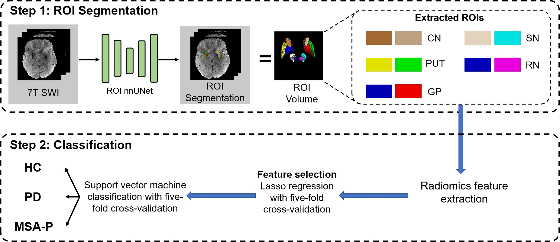
Figure 1 The workflow of radiomics analysis
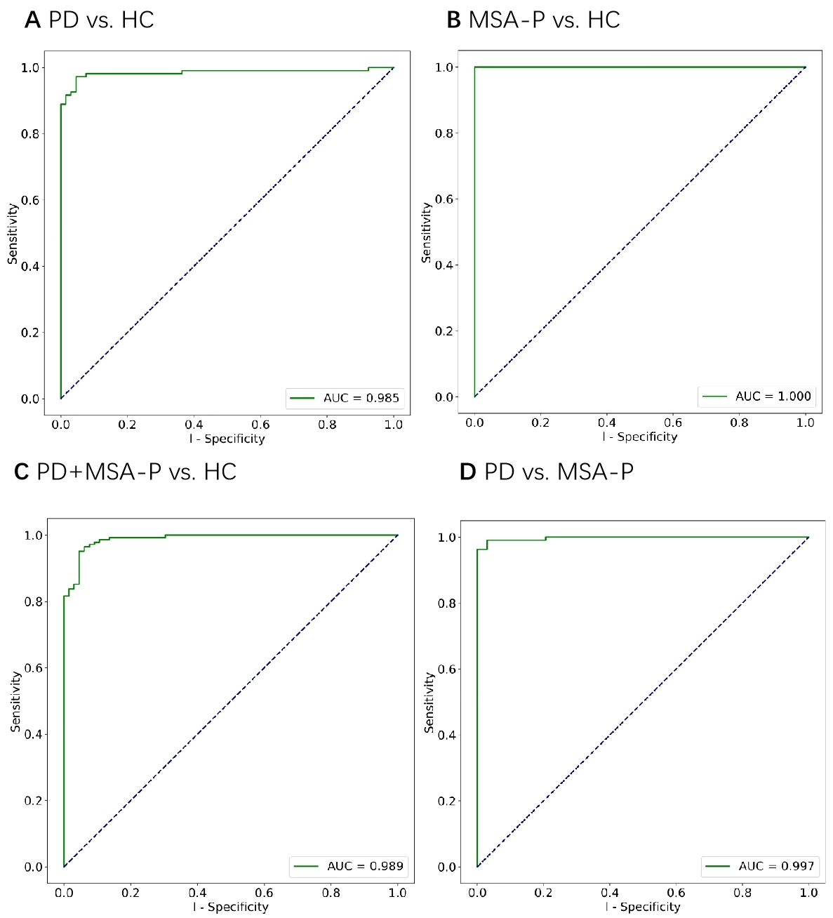
Figure 2 ROC curve analysis between groups
To cite this abstract in AMA style:
T. Feng, DN. Su, SD. Ding, ZJ. Zhang, Z. Zhang, J. Jing. Identification of Distinctive Iron Deposition Pattern in Subcortical Nuclei of Parkinson’s Disease using Radiomics: A 7T Susceptibility-Weighted Imaging Study [abstract]. Mov Disord. 2025; 40 (suppl 1). https://www.mdsabstracts.org/abstract/identification-of-distinctive-iron-deposition-pattern-in-subcortical-nuclei-of-parkinsons-disease-using-radiomics-a-7t-susceptibility-weighted-imaging-study/. Accessed April 7, 2026.« Back to 2025 International Congress
MDS Abstracts - https://www.mdsabstracts.org/abstract/identification-of-distinctive-iron-deposition-pattern-in-subcortical-nuclei-of-parkinsons-disease-using-radiomics-a-7t-susceptibility-weighted-imaging-study/