Category: Parkinson's disease: Neuroimaging
Objective: This study will investigate the relationships among speech-perception-in-noise (SPiN), hearing acuity, and cholinergic system changes in patients with PD.
Background: Recent studies highlight a critical gap in understanding of the associations between hearing sensitivity (e.g., pure-tone audiograms; PTA), hearing clarity (e.g., speech intelligibility in background noise), and cognitive decline in Parkinson’s disease (PD). Functional hearing loss, particularly impairments in speech perception in noise (SPiN), is prevalent in PD even with normal audiograms, suggesting central auditory processing deficits rather than cochlear synaptopathy or noise exposure as likely contributors. Emerging evidence implicates cholinergic system dysfunction, which plays a critical role not only in attention but also in auditory processing.
Method: 23 PD patients underwent QuickSIN SPiN, pure tone audiometry testing 1, and vesicular acetylcholine transporter (VAChT) [18F]FEOBV brain PET scans. Whole brain voxel-based analysis correlating VAChT binding with SPiN after adjustment for PTA was performed using SPM12.
Results: Voxel-based analysis showed significant cholinergic vulnerability in right temporo-occipital junctions, right superior temporal and temporal operculum auditory cortices, right dorsolateral prefrontal cortex, right orbitofrontal, posterior & anterior cingulum, right superior parietal cortex, right inferior colliculus, right superior and posterior cerebellar hemisphere, superior vermis, retrosplenial cortex, right occipital cortex (cluster-level FWER corrected P<0.05, see figure 1). A mask of statistically significant voxels was used to extract the mean [18F]FEOBV uptake value for regression analyses. A post-hoc regression analysis showed that greater central cholinergic system integrity associated with better SPiN performance (see figure 2) independently of pure tone audiometry (β=-0.507, p=0.013), accounting for an additional 25.4% of the variance in SPiN.
Conclusion: Greater cholinergic system integrity, predominantly lateralized to the right hemispheric auditory, lateral frontal, and multisensory cortices associated with better performance on the speech-perception-in-noise task. Prior works show that the right hemisphere appears to predominantly contribute to speech perception in noisy as compared to quiet conditions 2–4.
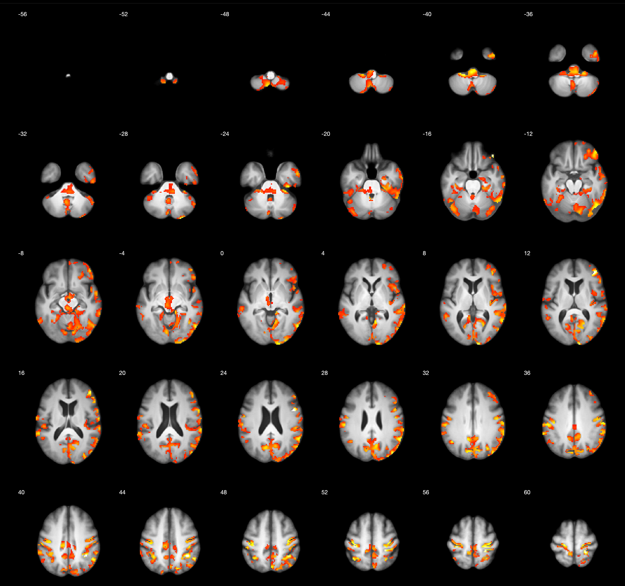
Figure 1.
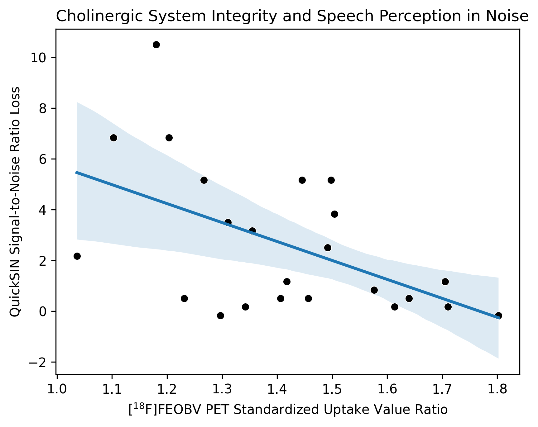
Figure 2.
References: 1. Fitzgerald, M. B., Gianakas, S. P., Qian, Z. J., Losorelli, S. & Swanson, A. C. Preliminary guidelines for replacing word-recognition in quiet with speech in noise assessment in the routine audiologic test battery. Ear Hear. 44, 1548–1561 (2023).
2. Shtyrov, Y. et al. Background acoustic noise and the hemispheric lateralization of speech processing in the human brain: magnetic mismatch negativity study. Neurosci. Lett. 251, 141–144 (1998).
3. Alexandrou, A. M., Saarinen, T., Mäkelä, S., Kujala, J. & Salmelin, R. The right hemisphere is highlighted in connected natural speech production and perception. Neuroimage 152, 628–638 (2017).
4. Yang, L. et al. Effects of age on the auditory cortex during speech perception in noise: Evidence from functional near-infrared spectroscopy. Ear Hear. 45, 742–752 (2024).
To cite this abstract in AMA style:
S. Roytman, D. Mccaslin, J. Barr, R. Vangel, P. Kanel, N. Bohnen. Impaired Speech Perception in Noise (SPiN), Hearing Loss, and Cholinergic Denervation in Parkinson’s Disease [abstract]. Mov Disord. 2025; 40 (suppl 1). https://www.mdsabstracts.org/abstract/impaired-speech-perception-in-noise-spin-hearing-loss-and-cholinergic-denervation-in-parkinsons-disease/. Accessed April 10, 2026.« Back to 2025 International Congress
MDS Abstracts - https://www.mdsabstracts.org/abstract/impaired-speech-perception-in-noise-spin-hearing-loss-and-cholinergic-denervation-in-parkinsons-disease/