Category: Parkinson's Disease: Neuroimaging
Objective: To characterize structural connectivity through whole-brain network-based statistics and graph theory implementation in treated Parkinson’s disease (PD) patients with probable REM sleep behavior disorder (RBD).
Background: Some studies have shown cognitive dysfunctions [1] and decreased white matter integrity measures in PD with RBD [2]. Alterations in white matter network has been previously explored in de novo PD probable RBD (pRBD) using graph theory showing changes in nodal efficiency mainly in neocortical and paralimbic regions [3]. However, as far we know there are no previous studies exploring whole-brain structural connectivity through network-based statistics in PD-pRBD.
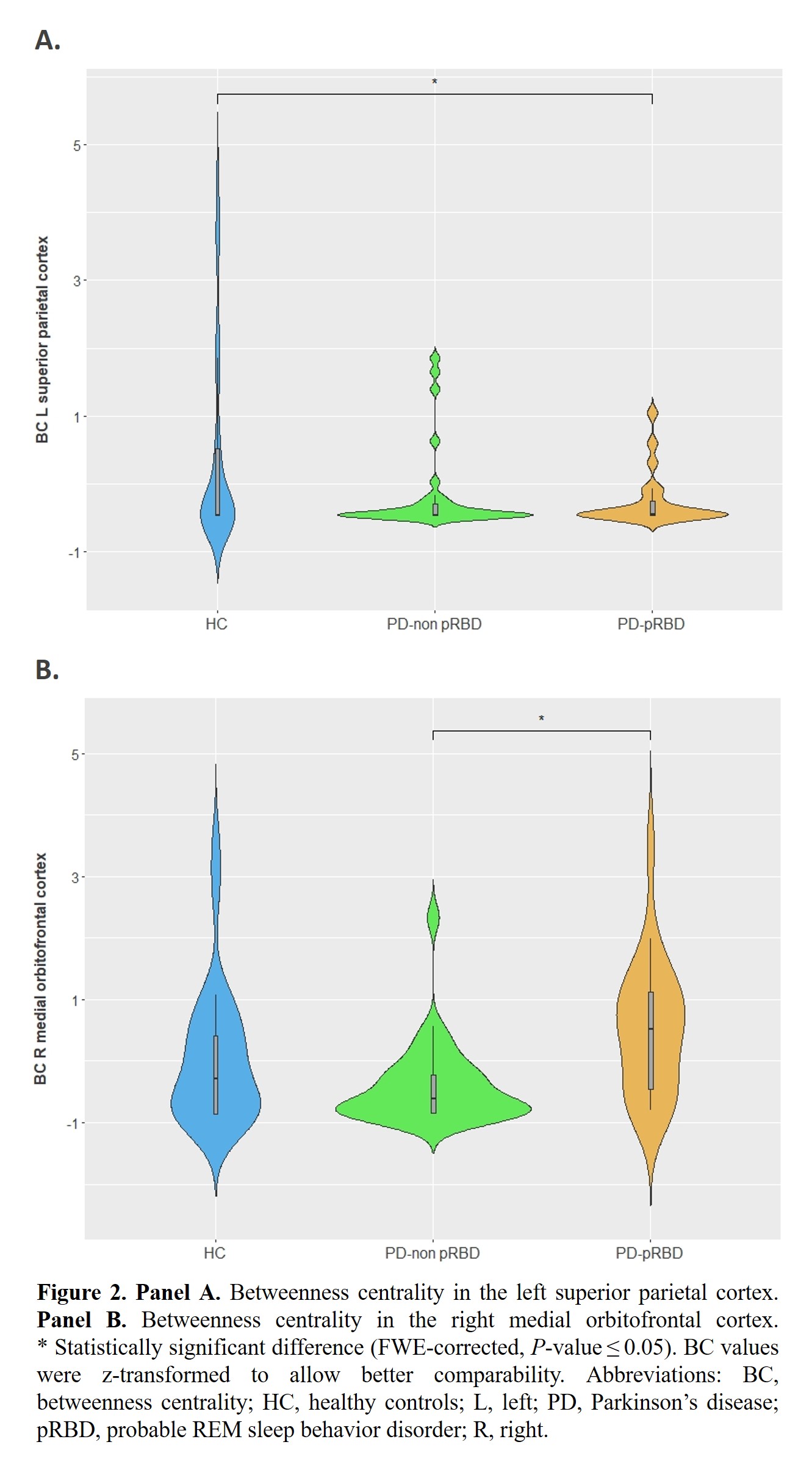
Method: The analyzed sample from the Hospital Clinic de Barcelona consisted of 22 PD-pRBD and 31 PD-non pRBD patients classified using the Innsbruck RBD Inventory [4], and 28 healthy controls [table1] who underwent diffusion-weighted 3T magnetic resonance imaging. Threshold-free network-based statistics was used to characterize interregional structural connectivity based on the number of streamlines (NOS) between 68 cortical [5] and 18 deep gray matter [6] regions of interest. Moreover, global and local measures of network integrity were computed using a graph theory implementation based on Brain Connectivity Toolbox [7]. Group comparisons included sex as a covariate.
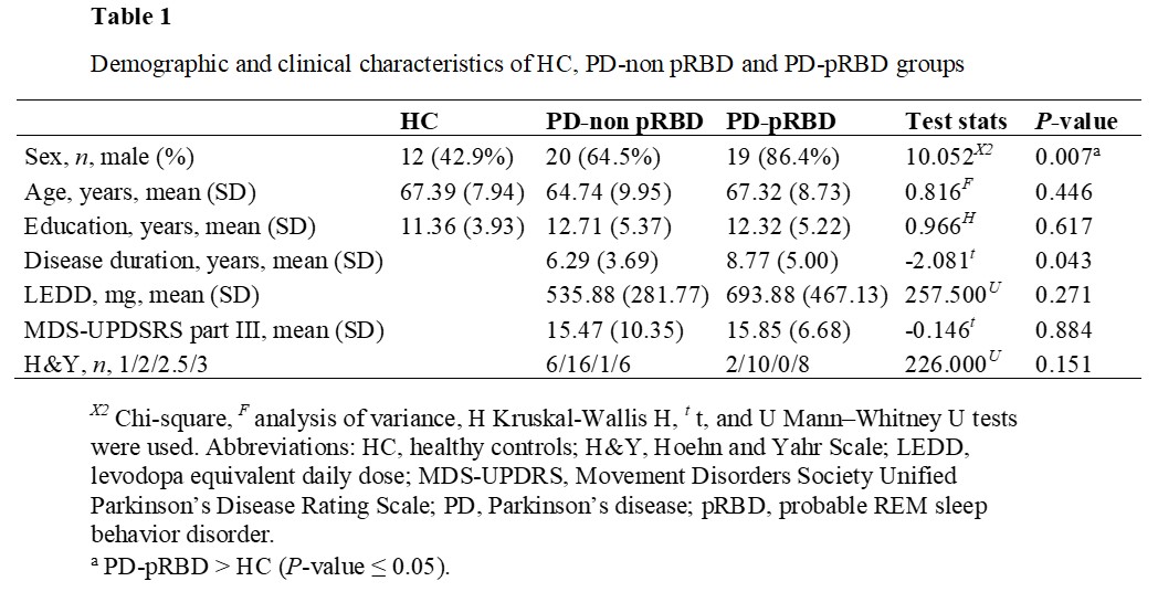
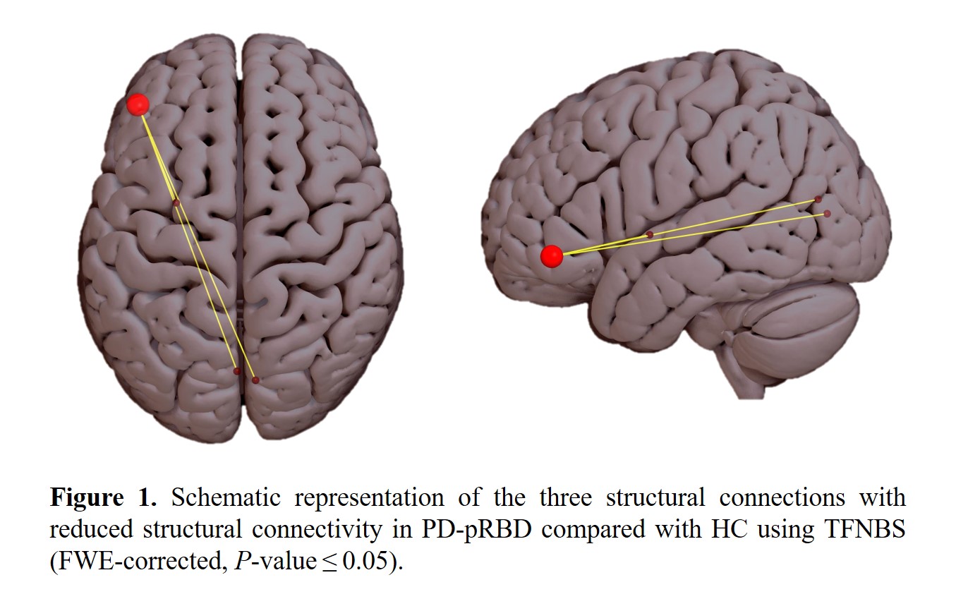
Results: PD-pRBD patients showed reduced structural connectivity compared with controls in three connections, involving left pars orbitalis’s connections with the left cuneus, left putamen, and right pericalcarine cortex (FWE corrected, P-value ≤ 0.05) [figure1]. Regarding graph measures, we found reduced betweenness centrality in the PD-pRBD group in the left superior parietal cortex compared with controls; and incremented betweenness centrality in the medial orbitofrontal cortex compared with the PD-non pRBD group (FWE corrected, P-value ≤ 0.05) [figure2a, figure2b], which remained significant after controlling for disease duration.
Conclusion: PD-pRBD patients show impairment of frontal pars orbitalis connectivity and a pattern of altered centrality in frontal and parietal regions.
*JO & IR contributed equally to this work as co-first authors.
References: 1. Mao J, Huang X, Yu J, et al. Association Between REM Sleep Behavior Disorder and Cognitive Dysfunctions in Parkinson’s Disease: A Systematic Review and Meta-Analysis of Observational Studies. Front Neurol. 2020;11:577874. Published 2020 Nov 6. doi:10.3389/fneur.2020.577874
2. Matzaras R, Shi K, Artemiadis A, et al. Brain Neuroimaging of Rapid Eye Movement Sleep Behavior Disorder in Parkinson’s Disease: A Systematic Review. J Parkinsons Dis. 2022;12(1):69-83. doi:10.3233/JPD-212571
3. Chen A, Li Y, Wang Z, et al. Disrupted Brain Structural Network Connection in de novo Parkinson’s Disease With Rapid Eye Movement Sleep Behavior Disorder. Front Hum Neurosci. 2022;16:902614. Published 2022 Jul 19. doi:10.3389/fnhum.2022.902614
4. Frauscher B, Ehrmann L, Zamarian L, et al. Validation of the Innsbruck REM sleep behavior disorder inventory. Mov Disord. 2012;27(13):1673-1678. doi:10.1002/mds.25223
5. Desikan RS, Ségonne F, Fischl B, et al. An automated labeling system for subdividing the human cerebral cortex on MRI scans into gyral based regions of interest. Neuroimage. 2006;31(3):968-980. doi:10.1016/j.neuroimage.2006.01.021
6. Fischl B, Salat DH, Busa E, et al. Whole brain segmentation: automated labeling of neuroanatomical structures in the human brain. Neuron. 2002;33(3):341-355. doi:10.1016/s0896-6273(02)00569-x
7. Rubinov M, Sporns O. Complex network measures of brain connectivity: uses and interpretations. Neuroimage. 2010;52(3):1059-1069. doi:10.1016/j.neuroimage.2009.10.003
To cite this abstract in AMA style:
J. Oltra, I. Roura, A. Campabadal, C. Uribe, J. Pardo, MJ. Martí, Y. Compta, F. Valldeoriola, N. Bargallo, A. Iranzo, C. Junque, B. Segura. Impaired structural connectivity in Parkinson’s disease with probable RBD [abstract]. Mov Disord. 2023; 38 (suppl 1). https://www.mdsabstracts.org/abstract/impaired-structural-connectivity-in-parkinsons-disease-with-probable-rbd/. Accessed June 4, 2026.« Back to 2023 International Congress
MDS Abstracts - https://www.mdsabstracts.org/abstract/impaired-structural-connectivity-in-parkinsons-disease-with-probable-rbd/