Category: Parkinson's Disease: Neuroimaging
Objective: We aimed to explore the relationship between the pNfL and change in the brain in early-stage PD using a longitudinal brain imaging cohort.
Background: Previous studies found that plasma neurofilament light protein (pNfL) was related to a greater rate of deterioration of motor and cognition in Parkinson’s disease (PD). However, the exact association between the pNfL and brain structure and function is still under research.
Method: Early-stage PD patients were recruited from the West China Hospital. All the patients received face-to-face interviews, blood sample collection, and brain magnetic resonance imaging (MRI) at baseline and follow-up. Gray matter (GM) volume and white matter volume were calculated as structure metrics. Fractional amplitude of low-frequency fluctuation (fALFF) and degree centrality (DC) were used to evaluate brain function. Patients were divided into the higher pNfL group and the lower pNfL group according to the median value of pNfL in the statistical analysis.
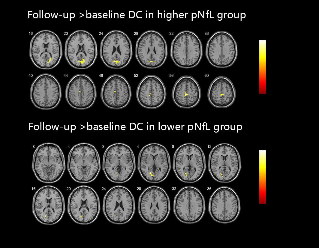
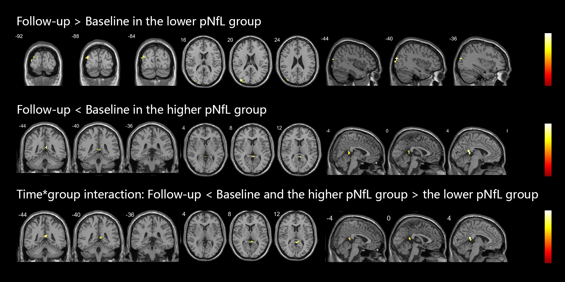
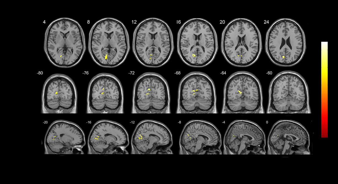
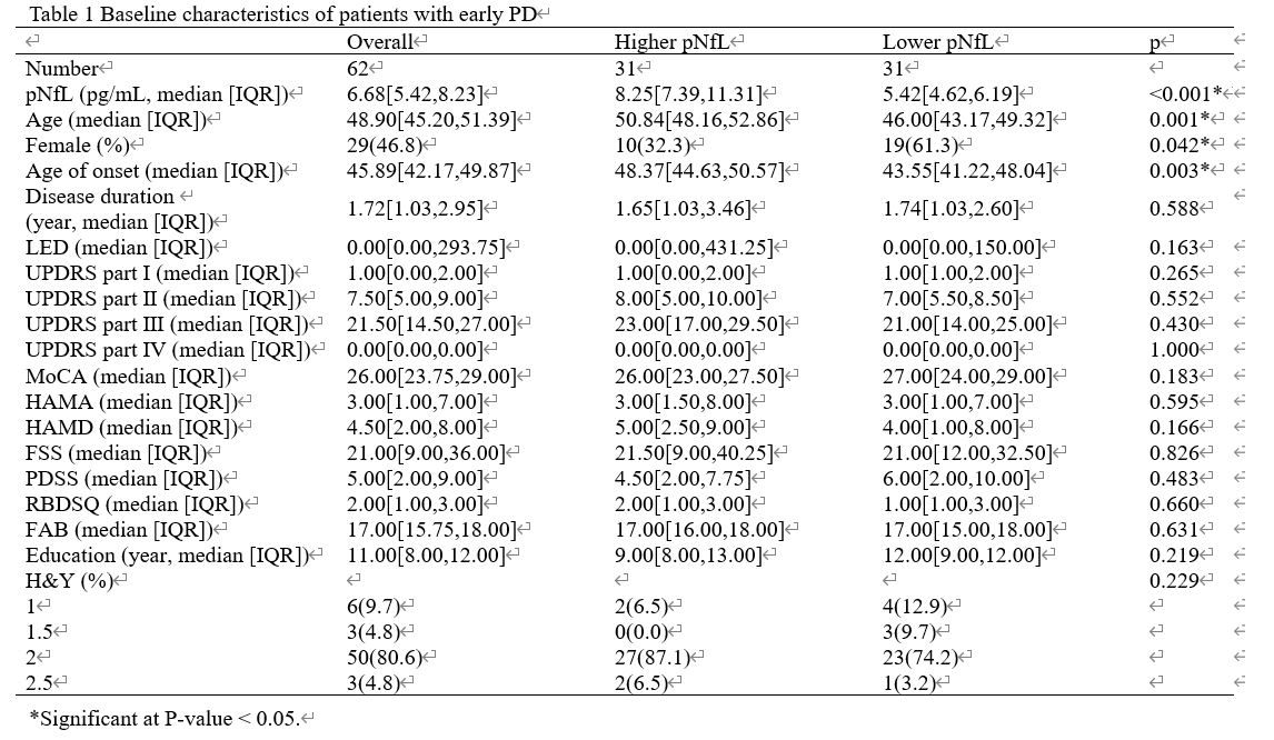
Results: There were 62 patients included in the current study, the baseline characteristics of them were listed in Table 1. We found that patients with higher pNfL had a faster decreased rate of fALFF in the right posterior cingulate compared to patients with lower pNfL indicated by the interaction between time and group (Figure 1). The higher pNfL group showed increased DC in the right calcarine and left paracentral lobule (Figure 2). The lower pNfL group only showed increased DC in the left superior occipital gyrus (Figure 2). In addition, a significantly decreased GM volume was found in the left calcarine at follow-up compared to that at baseline in the higher pNfL group (Figure 3).
Conclusion: Our study found that higher baseline pNfL predicted worse brain function and brain atrophy in the follow-up in patients with early-stage PD. Patients with higher baseline pNfL had a faster rate of decrease in regional brain function compared to those with lower pNfL. In addition, those with higher pNfL had significant GM atrophy in the follow-up, while those with lower pNfL had no detectable change in GM volume in a short follow-up period.
Figure 1 Longitudinal Change in fALFF in patients
Figure 2 Longitudinal Change in DC in patients
Figure 3 Longitudinal Change in GM in patients
Table 1 Demographic characteristics
To cite this abstract in AMA style:
Y. Xiao, SC. Wang, YB. Hou, HF. Shang. Longitudinal change in brain function and structure related to plasma neurofilament light protein in early-stage Parkinson’s disease [abstract]. Mov Disord. 2024; 39 (suppl 1). https://www.mdsabstracts.org/abstract/longitudinal-change-in-brain-function-and-structure-related-to-plasma-neurofilament-light-protein-in-early-stage-parkinsons-disease/. Accessed July 2, 2025.« Back to 2024 International Congress
MDS Abstracts - https://www.mdsabstracts.org/abstract/longitudinal-change-in-brain-function-and-structure-related-to-plasma-neurofilament-light-protein-in-early-stage-parkinsons-disease/


