Objective: To identify distinct subtypes of Parkinson’s disease (PD) based on diffusion tensor imaging (DTI)-derived free water (FW) patterns in basal ganglia (BG) structures using machine learning (ML) and to examine their relationship with motor symptom severity and progression over two years.
Background: PD is a highly heterogeneous neurodegenerative disorder characterized by different subtypes (1). Brain atrophy in PD leads to increased FW accumulation (2), which can be quantified using isotropic diffusion (ISO), a DTI-derived measure (3). The BG, a key structure affected in PD, may exhibit heterogeneous FW distribution patterns that correlate with motor symptom severity. This study leveraged unsupervised ML to identify PD subtypes based on ISO values and assess their motor progression over two years.
Method: Baseline DTI and MDS-UPDRS-III motor scores (at baseline and 2-year follow-up) from 156 PD patients were obtained from the PPMI database (4). ISO values were extracted from the bilateral Red Nucleus, Substantia Nigra, Subthalamic Nucleus, Striatum, and both Globus Pallidus Externus and Internus using the BG Atlas in DSI Studio (5). These ISO values were used as input for hierarchical clustering to identify patient subtypes. Motor symptom severity between subtypes was compared using two sample t-tests. A 2-year follow-up analysis was conducted in 95 medication-free patients to assess motor symptom progression.
Results: Clustering analysis revealed two distinct PD subtypes. Subtype 2 exhibited significantly higher ISO values across all BG white matter tracts compared to Subtype 1 (p < 0.001; Figure 1). Clinically, Subtype 2 showed significantly increased bradykinesia and rigidity scores (p < 0.001; Figure 2). These findings were confirmed in the 2-year follow-up cohort (n=95), where Subtype 2 continued to demonstrate worse motor symptom progression compared to Subtype 1 (Figure 3).
Conclusion: Our study reveals distinct PD subtypes characterized by different patterns of FW accumulation in BG structures, with higher ISO values associated with more severe motor symptoms. The persistence of these patterns over two years suggests these represent stable disease subtypes rather than temporal variations. Future research should explore the underlying mechanisms of these differences and their implications for personalized treatment strategies.
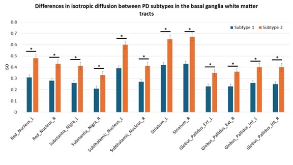
Differences in ISO
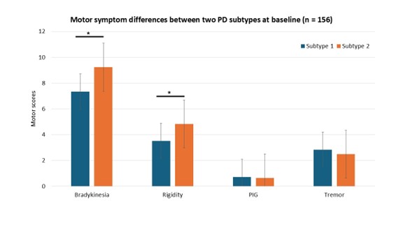
Motor Score Differences at Baseline
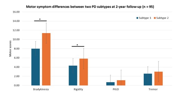
Motor Score Differences at the 2-Year Mark
References: 1. Vijayakumari AA, Fernandez H, Walter B. Data-driven Approach to Identify Subtypes and Progression of Parkinson’s disease using Multimodal Imaging Data (P6-11.006). Neurology. 2023;100(17 Supplement 2):1311.
2. Andica C, Kamagata K, Hatano T, Saito A, Uchida W, Ogawa T, et al. Free-Water Imaging in White and Gray Matter in Parkinson’s Disease. Cells. 2019;8(8).
3. Bower AE, Crisomia SJ, Chung JW, Martello JP, Burciu RG. Free water imaging unravels unique patterns of longitudinal structural brain changes in Parkinson’s disease subtypes. Front Neurol. 2023;14:1278065.
4. Marek K, Chowdhury S, Siderowf A, Lasch S, Coffey CS, Caspell-Garcia C, et al. The Parkinson’s progression markers initiative (PPMI) – establishing a PD biomarker cohort. Ann Clin Transl Neurol. 2018;5(12):1460-77.
5. Yeh FC, Wedeen VJ, Tseng WY. Generalized q-sampling imaging. IEEE Trans Med Imaging. 2010;29(9):1626-35.
To cite this abstract in AMA style:
A. Vijayakumari, H. Fernandez, B. Walter. Machine Learning Analysis of DTI-Derived Free Water Reveals Distinct Parkinson’s Disease Subtypes: A Two-Year Longitudinal Study [abstract]. Mov Disord. 2025; 40 (suppl 1). https://www.mdsabstracts.org/abstract/machine-learning-analysis-of-dti-derived-free-water-reveals-distinct-parkinsons-disease-subtypes-a-two-year-longitudinal-study/. Accessed April 7, 2026.« Back to 2025 International Congress
MDS Abstracts - https://www.mdsabstracts.org/abstract/machine-learning-analysis-of-dti-derived-free-water-reveals-distinct-parkinsons-disease-subtypes-a-two-year-longitudinal-study/