Category: Rare Neurometabolic Diseases
Objective: To evaluate microstructural changes in the dorsal striatum and their correlation with clinical symptoms in Wilson disease (WD).
Background: Advanced diffusion models, including Neurite Orientation Dispersion and Density Imaging (NODDI), offer a sophisticated approach for assessing the microstructural complexity of brain tissue from diffusion-weighted magnetic resonance imaging (dMRI) by quantifying neurite morphology [1]. To date, two studies have identified NODDI changes in the striatum in WD [2,3]; however, their relationship with cognitive function has yet to be investigated.
Method: 19 WD patients and 19 age- and sex-matched healthy controls underwent 3T multi-shell dMRI and were evaluated using the Unified WD Rating Scale neurological subscale (UWDRS-N) and the Montreal Cognitive Assessment (MoCA) [Table 1]. The CUDA diffusion modelling toolbox (cuDIMOT [4]) of the FMRIB Software Library (FSL) was employed to estimate the intra-cellular volume fraction (Vic; indicating neurite density), orientation dispersion (OD; indicating axonal and dendritic dispersion), and isotropic volume fraction (Viso; indicating cerebrospinal fluid) of the NODDI model [1]. The processing steps for dMRI data are summarized in [Figure 1]. Diffusion metrics were compared between groups using Mann-Whitney U tests, and their associations with clinical scores were explored using Spearman’s correlations in SPSS Statistics. A Bonferroni-corrected p ≤ .05 was considered significant.
Results: The striatum of WD patients exhibited significantly higher Vic and Viso, as well as lower OD, when compared to controls [Figure 2]. UWDRS-N scores were positively correlated with the Viso (rs = .40, p = .036; rs = .55, p = .001) and negatively correlated with the OD of the putamen and caudate nucleus (rs = -.43, p = .022; rs = -.59, p < .001). MoCA scores were negatively correlated with the putaminal Viso (rs = -.44, p = .017).
Conclusion: Consistent with prior research [2,3], we demonstrated increased Viso and diminished OD in the striatum of WD patients, which may reflect axonal loss, myelin damage, decreased dendrite density, and spongiform degeneration. The elevated Vic in patients may indicate a compensatory mechanism that increases neurite density under decoppering treatment. Increased Viso of the putamen was correlated with greater neurological and cognitive impairment, suggesting it may serve as a valuable biomarker for clinical symptoms in WD.
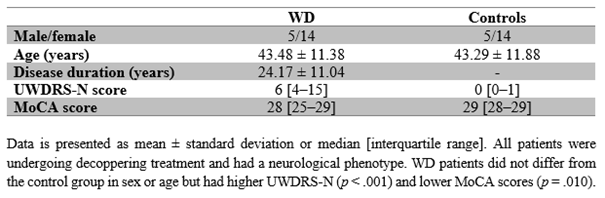
Table 1. Demographics and Clinical Scores
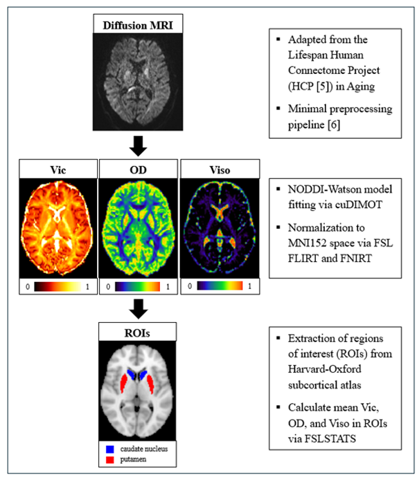
Figure 1. Processing Steps for the dMRI Analysis
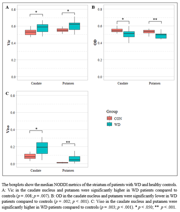
Figure 2. Intergroup Differences in NODDI Metrics
References: [1] Zhang, H., Schneider, T., Wheeler-Kingshott, C. A., & Alexander, D. C. (2012). NODDI: practical in vivo neurite orientation dispersion and density imaging of the human brain. Neuroimage, 61(4), 1000-1016. doi: 10.1016/j.neuroimage.2012.03.072
[2] Song, Y., Li, X., Huang, X., et al. (2018). A study of neurite orientation dispersion and density imaging in Wilson’s disease. International Society for Magnetic Resonance in Medicine, 48, 423-430. doi: 10.1002/jmri.25930
[3] Su, D., Zhang, Z., Zhang, Z., et al. (2023). Microstructural and functional impairment of the basal ganglia in Wilson’s disease: A multimodal neuroimaging study. Frontiers in Neuroscience, 17, 1146644. doi: 10.3389/fnins.2023.1146644
[4] Hernandez-Fernandez, M., Reguly, I., Jbabdi, S., et al. (2019). Using GPUs to accelerate computational diffusion MRI: From microstructure estimation to tractography and connectomes. Neuroimage, 188, 598-615. doi: 10.1016/j.neuroimage.2018.12.015
[5] Harms, M. P., Somerville, L. H., Ances, B. M., et al. (2018). Extending the Human Connectome Project across ages: Imaging protocols for lifespan development and aging projects. Neuroimage, 183, 972-984. doi: 10.1016/j.neuroimage.2018.09.060
[6] Glasser, M. F., Sotiropoulos, S. N., Wilson, A., et al. (2013). The minimal preprocessing pipelines for the Human Connectome Project. Neuroimage, 80, 105-124. doi: 10.1016/j.neuroimage.2013.04.127
To cite this abstract in AMA style:
A. Hausmann, S. Querbach, C. Rubbert, A. Schnitzler, J. Caspers, C. Hartmann. Microstructural Changes in the Striatum Correlate With Clinical Symptoms in Wilson Disease [abstract]. Mov Disord. 2025; 40 (suppl 1). https://www.mdsabstracts.org/abstract/microstructural-changes-in-the-striatum-correlate-with-clinical-symptoms-in-wilson-disease/. Accessed April 10, 2026.« Back to 2025 International Congress
MDS Abstracts - https://www.mdsabstracts.org/abstract/microstructural-changes-in-the-striatum-correlate-with-clinical-symptoms-in-wilson-disease/