Category: Parkinson's Disease: Pathophysiology
Objective: To define the profile of different cell signaling molecules in the blood of patients with Parkinson’s Disease (PD) compared to controls.
Background: PD is one of the most prevalent neurodegenerative disorders, characterized by debilitating motor impairments and cognitive symptoms. The diagnosis of PD primarily relies on clinical assessment, which typically occurs after significant neural damage. Although efforts have been made to identify effective biomarkers for early diagnosis of PD, none currently provide conclusive results [1]. Various cell signaling molecules are relevant in the context of neurodegenerative diseases. Nevertheless, conflicting findings persist, emphasizing the urgent need for further research.
Method: We enrolled 24 patients with PD and 20 age- and gender-matched controls. On the same day of the clinical assessment, blood was collected and processed for plasma separation. Using ELISA or Multiplex assay, we evaluated the plasma levels of Brain-Derived Neurotrophic Factor (BDNF), Platelet-Derived Growth Factor (PDGF) with subunits AA and AB/BB, Soluble Intercellular Adhesion Molecule-1 (sICAM-1), Soluble Vascular Cell Adhesion Molecule-1 (sVCAM-1), Neural Cell Adhesion Molecule (NCAM), Plasminogen Activator Inhibitor-1 (PAI-1), Cathepsin D, and Regulated on Activation Normal T Cell Expressed and Secreted (RANTES).
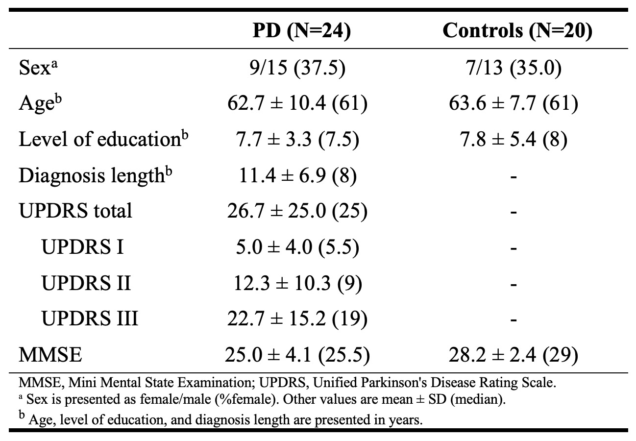
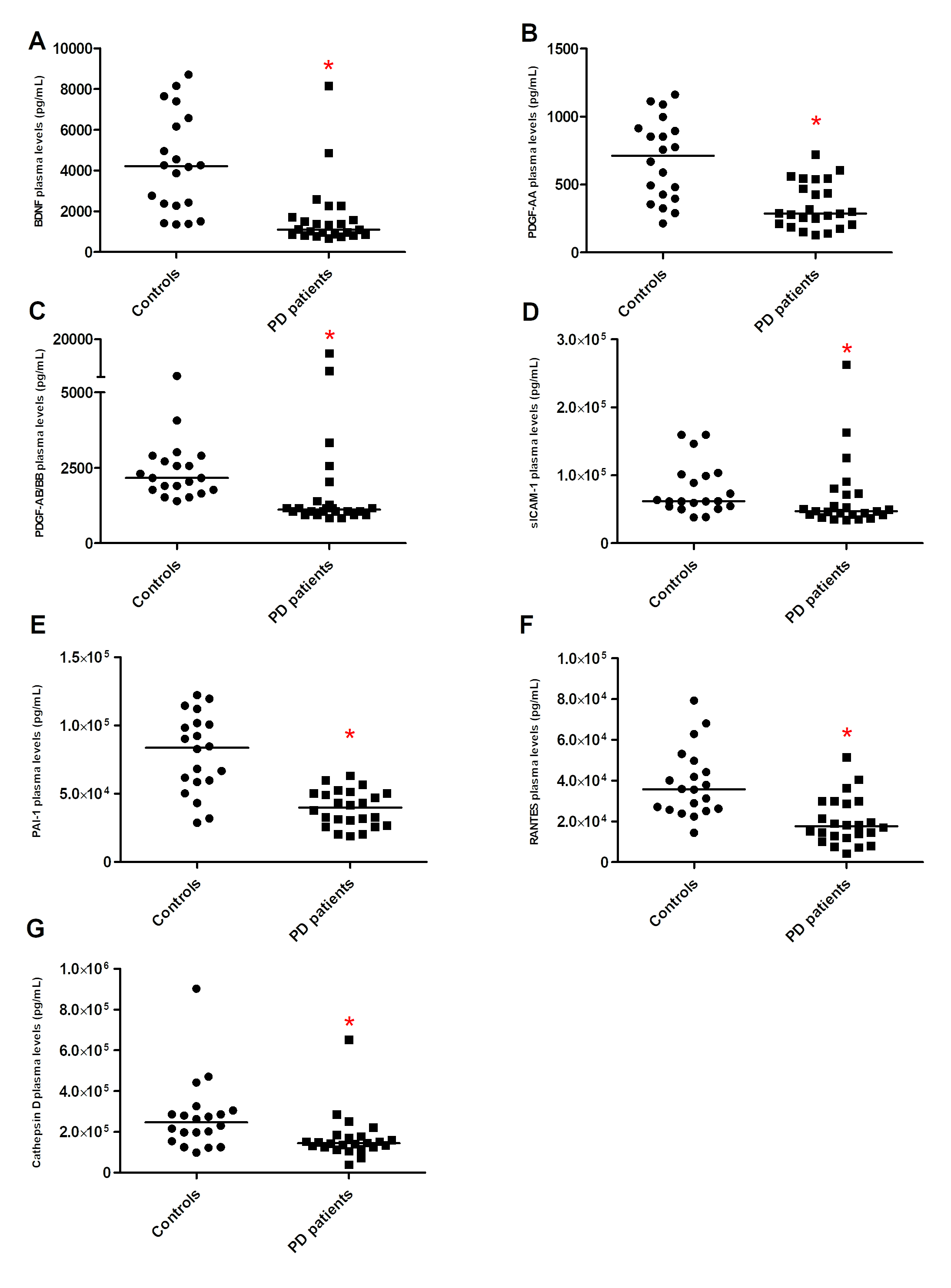
Results: A general description of the study population is provided in table 1. The plasma levels of the growth factors, including BDNF, PDGF-AA and PDGF-AB/BB, were significantly lower in PD patients than controls (BDNF: 1,690 vs. 4,309 pg/mL, p <0.0001; PDGF-AA: 346 vs. 683 pg/mL, p = 0.0002; PDGF-AB/BB: 2,084 vs. 2,415 pg/mL, p = 0.0002) [figure 1]. Among the four cell adhesion molecules, two showed a significant decrease in PD patients compared to the control group (sICAM-1: 67,192 vs. 79,418 pg/mL, p = 0.0196; PAI-1: 39,331 vs. 79,458 pg/mL, p <0.0001; sVCAM-1: 1,003 vs. 1,103 ng/mL, p = 0.7681; NCAM: 331,554 vs. 326,145 pg/mL, p = 0.8136). Patients with PD showed a significantly lower plasma levels of the Cathepsin D (170,530 vs. 275,712 pg/mL, p = 0.0039) and the chemokine, RANTES (20,036 vs. 38,705 pg/mL, p = 0.0002).
Conclusion: Patients with PD exhibited reduced plasma levels of various cell signaling molecules compared to controls. Future longitudinal studies are warranted to investigate the dynamic changes of these markers over time and in different stages of PD.
Table 1. Study population
Figure 1. Cell signaling molecules plasma levels
References: 1. Tönges L, Buhmann C, Klebe S, Klucken J, Kwon EH, Müller T, Pedrosa DJ, Schröter N, Riederer P, Lingor P. Blood-based biomarker in Parkinson’s disease: potential for future applications in clinical research and practice. J Neural Transm (Vienna). 2022 Sep;129(9):1201-1217.
To cite this abstract in AMA style:
S. Zadegan, A. Teixeira, E. Furr Stimming, N. Pessoa Rocha. Reduced plasma levels of various cell signaling molecules in patients with Parkinson’s Disease [abstract]. Mov Disord. 2024; 39 (suppl 1). https://www.mdsabstracts.org/abstract/reduced-plasma-levels-of-various-cell-signaling-molecules-in-patients-with-parkinsons-disease/. Accessed July 3, 2025.« Back to 2024 International Congress
MDS Abstracts - https://www.mdsabstracts.org/abstract/reduced-plasma-levels-of-various-cell-signaling-molecules-in-patients-with-parkinsons-disease/


