Category: Neuroimaging (Non-PD)
Objective: Ex vivo 7T MRI can identify macrostructural changes in the basal ganglia that reflect underlying neuropathology, and can be used to differentiate between proteinopathies causing clinical frontotemporal lobar dementia syndromes (FTD).
Background: Accurate prediction of underlying neuropathology based on clinical phenotype poses a significant challenge to the development of therapeutics in FTD. Frontotemporal lobar degeneration (FTLD) due to TDP-43 (FTLD-TDP) vs tau (FTLD-Tau) can be clinically indistinguishable during life. Ex vivo 7T MRI allows ultra-high resolution imaging of structure and is particularly sensitive to iron, which accumulates in activated glia in the cortex of FTLD but is understudied in the basal ganglia.
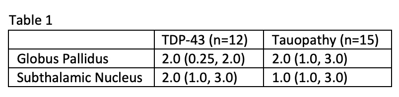
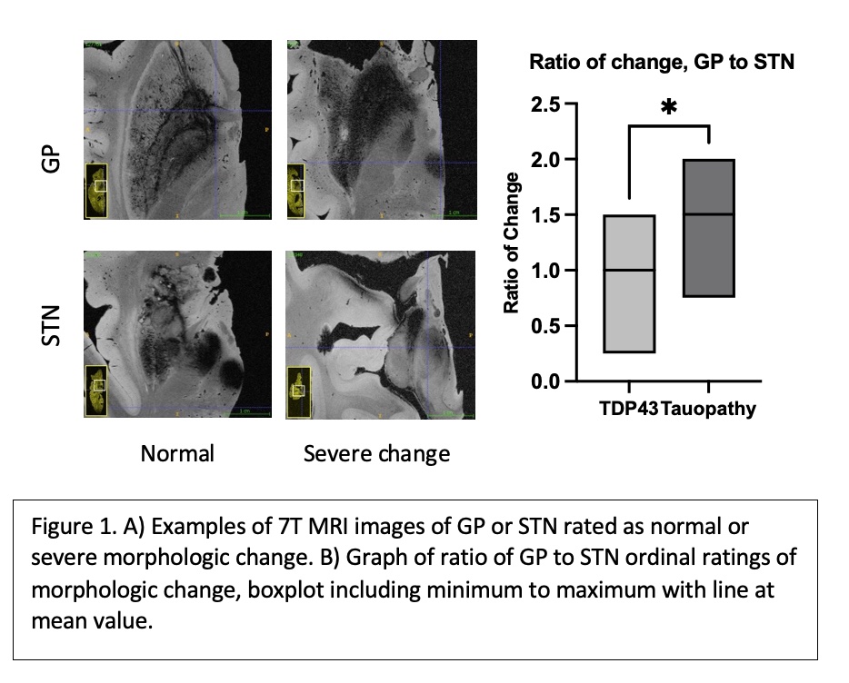
Method: A cohort of patients with clinical FTD underwent autopsy in which one hemisphere was taken for neuropathologic analysis, while the contralateral hemisphere was imaged at 160 µm isotropic resolution with a T2*w gradient-echo sequence using a whole-body 7T scanner (MAGNETOM Terra, Siemens Healthineers, Erlangen, Germany). MRI images were systematically reviewed, blinded to neuropathologic diagnosis to identify radiographic features of the basal ganglia which were altered in structure and/or intensity compared to control hemispheres. We identified 2 structural features of interest and developed a 4 point ordinal scale (i.e. none, mild, moderate severe) for the loss of normal morphological boundaries in globus pallidus (GP) and subthalamic nucleus (STN).
Results: FTLD-TDP43 group trended towards diminished STN radiographic morphology and relative preservation of GP compared with FTLD-Tau on ex vivo 7T2*w MRI (see [table1] reporting median ordinal rating and interquartile range). Overall, a ratio of change in morphological boundary of GP to STN was significantly different between the two groups [figure1].
Conclusion: Ultra-high resolution ex vivo 7T MRI reveals loss of structural integrity of the GP with preserved STN morphology at a radiographic level in brains with accumulation of tau compared to TDP43, which may aid in more accurate diagnosis of FTLD neuropathology in vivo.
To cite this abstract in AMA style:
R. Williamson, P. Khandelwal, S. Das, G. Mizsei, K. Prabhakaran, R. Ittyerah, D. Wolk, J. Detre, P. Yushkevich, E. Lee, J. Gee, M. Grossman, C. Mcmillan, M. Tisdall, D. Irwin. Structural analysis of the basal ganglia using ex vivo 7T MRI can differentiate frontotemporal lobar degeneration with TDP-43 vs tau accumulation. [abstract]. Mov Disord. 2023; 38 (suppl 1). https://www.mdsabstracts.org/abstract/structural-analysis-of-the-basal-ganglia-using-ex-vivo-7t-mri-can-differentiate-frontotemporal-lobar-degeneration-with-tdp-43-vs-tau-accumulation/. Accessed May 24, 2026.« Back to 2023 International Congress
MDS Abstracts - https://www.mdsabstracts.org/abstract/structural-analysis-of-the-basal-ganglia-using-ex-vivo-7t-mri-can-differentiate-frontotemporal-lobar-degeneration-with-tdp-43-vs-tau-accumulation/