Session Information
Date: Wednesday, June 22, 2016
Session Title: Huntington's disease
Session Time: 12:00pm-1:30pm
Location: Exhibit Hall located in Hall B, Level 2
Objective: To assess feasibility of measuring pathological tau deposition in patients with Huntington’s disease (HD) using [18F]-AV-1451 (f.k.a. T807) PET.
Background: HD is a progressive neurodegenerative disorder associated with pathological accumulation of the Huntington’s protein caused by more than 35 CAG triplet repeats within the HTT gene. Recently, the microtubule associated protein tau, which has previously been implicated as a hallmark of several other neurodegenerative disorders, has been proposed as a potential disease-modifying factor in HD. Recent post mortem investigations reported elevated tau levels primarily in the putamen of HD patients (Vuono et al., 2015). [18F]-AV-1451, a new tracer reliably binding to the pathological aggregates of tau offers the opportunity to examine tau pathology in vivo (Marquié et al., 2015). Therefore, our current study aims to assess tau pathology in living HD patients using [18F]-AV-1451-PET.
Methods: We acquired PET images from three stage II HD patients (mean age = 64± 2) using [18F]-AV-1451. After standard image preprocessing and intensity standardization, we calculated the Z-transformed tracer uptake deviation between HD patients and the mean uptake of an age-matched healthy control cohort (n = 16; mean age = 62±10).
Results: We report areas exceeding mean tau deposition in healthy controls by at least 2 standard deviations (z > 2) (see Figure 1): Patient 1 and 2 showed greatest pathological tau accumulation (z > 4) within the basal ganglia, peaking in the right pallidum as well as focal tau pathology in the cerebellum. Regarding the cerebral cortex, patient 2 showed increased tau accumulation in the right occipital lobe, patient 1 in temporal and parietal lobes bilaterally. In contrast, patient 3 displayed greatest (z > 4) and most spatially extended tau signal deviation within the anterior cerebellum bilaterally and moderate tau accumulation within the right hippocampus. 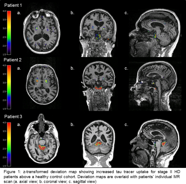
Conclusions: Here we demonstrate the feasibility of measuring increased tau deposition in three HD patients using [18F]-AV-1451-PET. Tau tracer uptake considerably varied between patients, but was most pronounced in the putamen and cerebellum. These results corroborate recent neuropathological findings and underscore the implication of tau pathology in HD.
To cite this abstract in AMA style:
K. Giehl, K. Reetz, I. Dogan, C. Werner, J.B. Schulz, A. Drzezga, T. van Eimeren. Tau pathology in Huntington’s disease: A brief in vivo PET-imaging report [abstract]. Mov Disord. 2016; 31 (suppl 2). https://www.mdsabstracts.org/abstract/tau-pathology-in-huntingtons-disease-a-brief-in-vivo-pet-imaging-report/. Accessed March 23, 2026.« Back to 2016 International Congress
MDS Abstracts - https://www.mdsabstracts.org/abstract/tau-pathology-in-huntingtons-disease-a-brief-in-vivo-pet-imaging-report/