Session Information
Date: Thursday, June 8, 2017
Session Title: Parkinson's Disease: Neuroimaging And Neurophysiology
Session Time: 1:15pm-2:45pm
Location: Exhibit Hall C
Objective: Appraise how cognitive performance and neural response is affected by nigrostriatal denervation in early-stage Parkinson’s disease (PD).
Background: Cognitive dysfunction in PD is heterogeneous and relates in part to nigrostriatal dopaminergic denervation 1. Resting state functional magnetic resonance imaging (rs-fMRI) is a non-invasive method to assess grey matter connectivity (rs-FC) and can be used to identify cognitive networks in PD. We hypothesized that reduction in striatal dopamine and cognitive dysfunction, individually and in combination, would influence connectivity within select cortico-striatal networks.
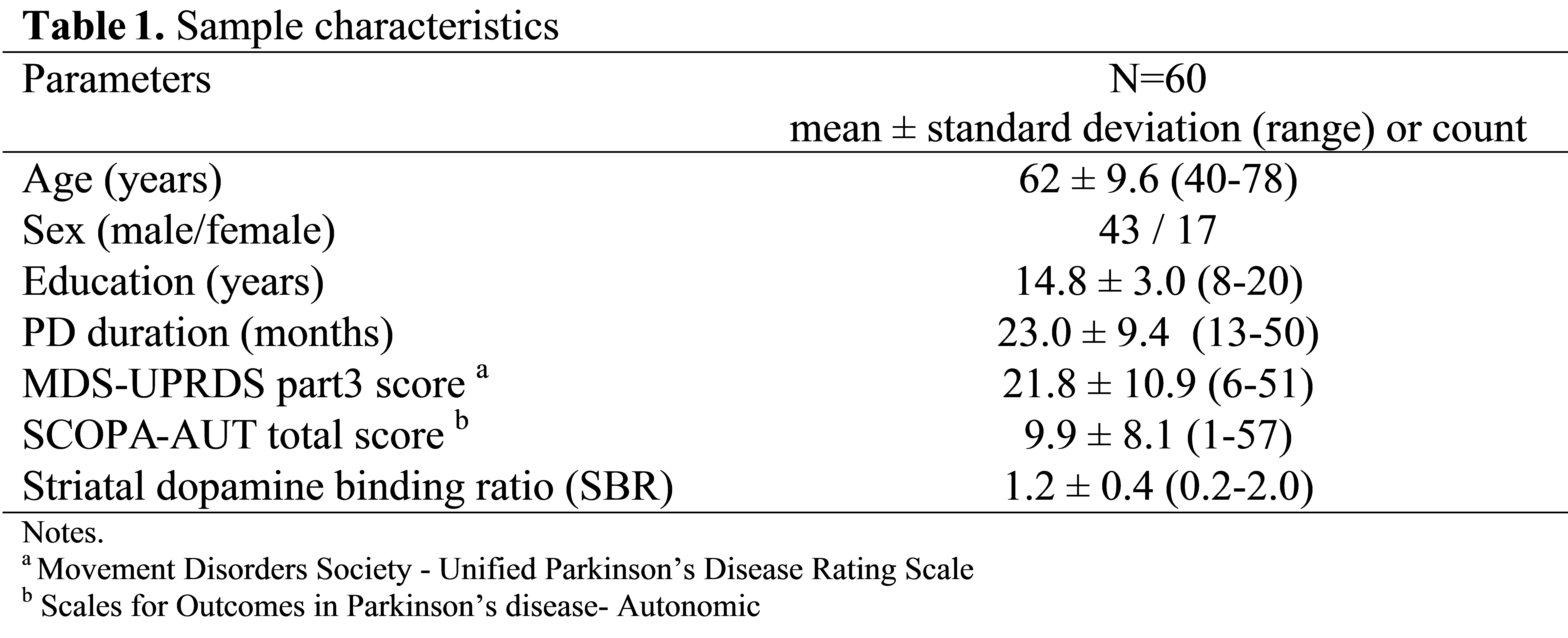
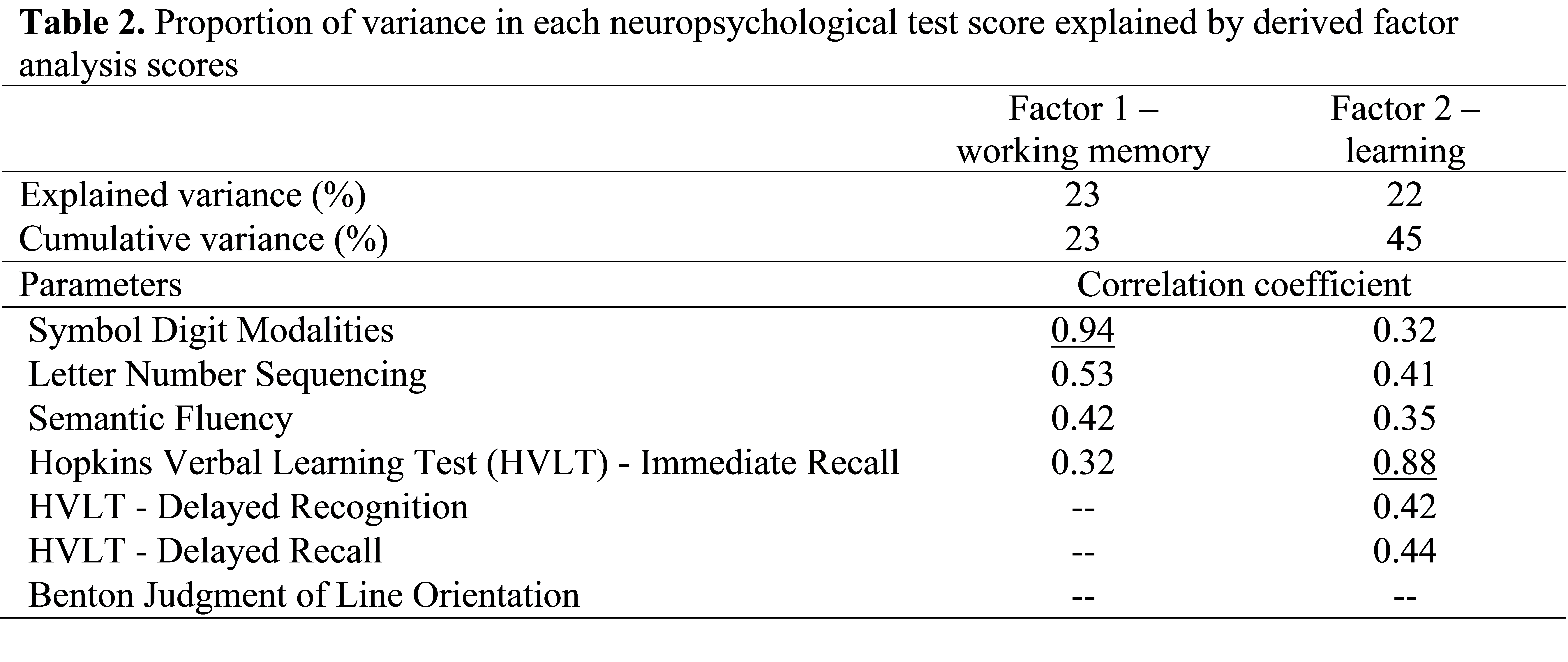
Methods: Sixty participants (Mage=61.8±9.6 years; Mduration=23±9.4 months) from the Parkinson’s Progression Markers Initiative (PPMI) were selected for clinical diagnosis of PD, rs-fMRI and 123I-FP-CIT SPECT scans, and excluded for absent dopaminergic deficits (SWEDDS) [table1]. Factor analysis reduced neuropsychological data into two latent cognitive factors [table2]. Mean striatal dopamine binding ratio (SBR) was derived from dopamine transporter SPECT of bilateral caudate and putamen. Bilateral caudate and putamen (Harvard-Oxford atlas) were used to create two seeds for rs-fMRI analysis (CONNtoolbox-15g). Two whole-brain general linear models (1 for each cognitive factor) controlling for age and education tested main effects of cognitive factor, SBR, and interaction (p<.001, cluster corrected p<.05).
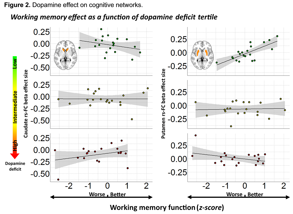
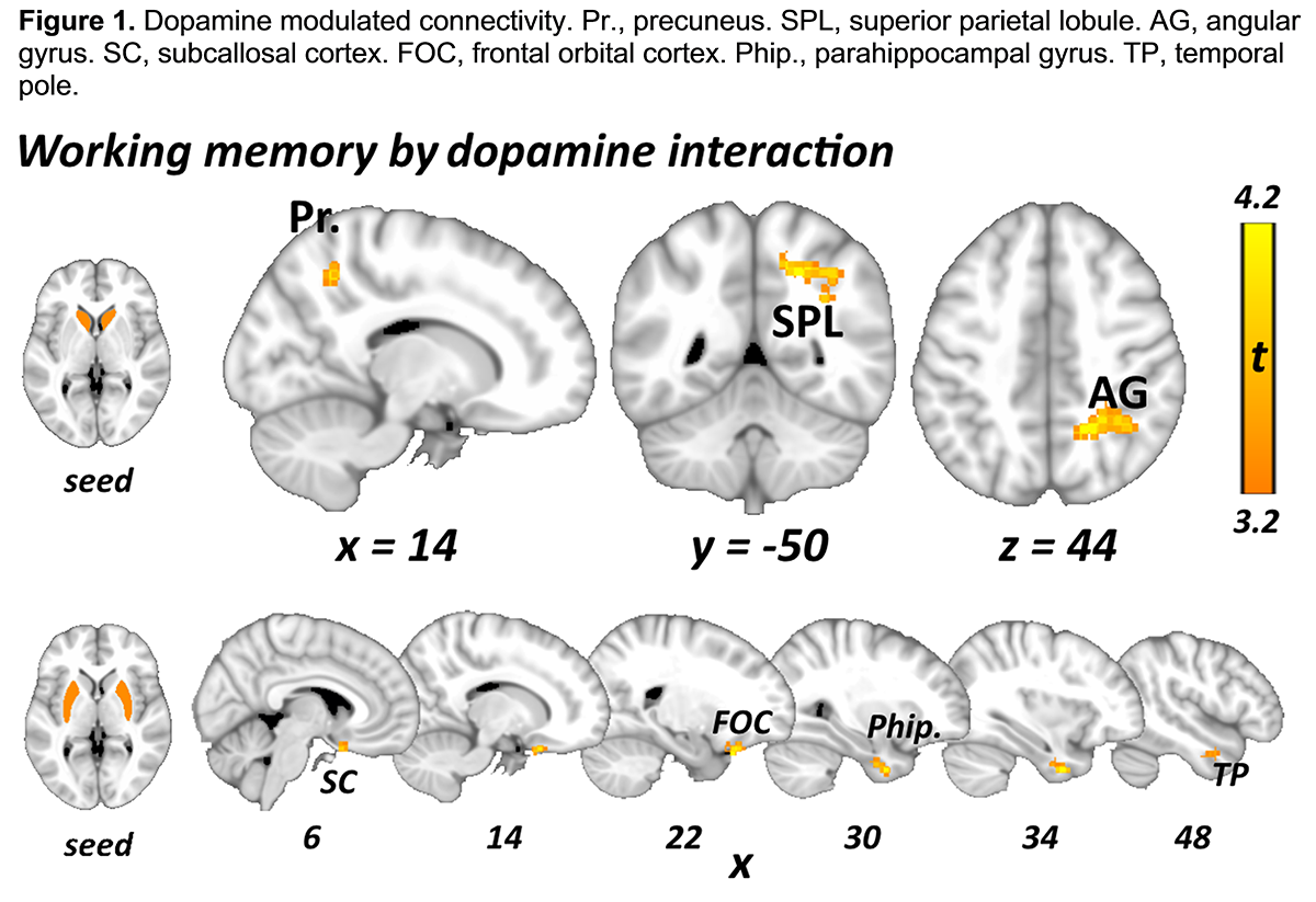
Results: No main effects of cognitive factor or SBR were found. Factor 1 (“working memory”) by SBR interaction was found for caudate to a cluster in the right precuneus-angular gyrus (Pr-AG), and for putamen to a cluster in the orbitofrontal cortex (OFC) [figure1]. Visualization plots demonstrated that the interaction resulted from a U-shaped dopamine-connectivity response curve mapping onto working memory function [figure2]. No interaction for Factor 2 (“learning”) was found.
Conclusions: Caudate-default mode network (Pr-AG) & putamen-salience network (OFC) connectivity showed opposing interactions. U-shaped functions in dopamine-brain response are well described in animal models, but, to our knowledge this is the first description relating to working memory in PD using rs-FC. Opposing interactions may be explained by different roles of default mode and salience networks in cognition. Network rs-FC may explain preserved performance in deficit.
References: (1) Lebedev A V, Westman E, Simmons A, et al. Large-scale resting state network correlates of cognitive impairment in Parkinson’s disease and related dopaminergic deficits. Front Syst Neurosci. 2014;8:45. doi:10.3389/fnsys.2014.00045.
An earlier version of this research was presented at the Alzheimer’s Association International Conference (07/2016, Toronto, ON).
To cite this abstract in AMA style:
A. Metcalfe, S. Udow, H. Sharmarke, Z. Shirzadi, D. Long, S. Duff-Canning, C. Marras, A. Lang, B. MacIntosh, M. Masellis. U-shaped dopamine resting state connectivity response function related to working memory performance in adults with early-stage Parkinson’s disease [abstract]. Mov Disord. 2017; 32 (suppl 2). https://www.mdsabstracts.org/abstract/u-shaped-dopamine-resting-state-connectivity-response-function-related-to-working-memory-performance-in-adults-with-early-stage-parkinsons-disease/. Accessed July 1, 2025.« Back to 2017 International Congress
MDS Abstracts - https://www.mdsabstracts.org/abstract/u-shaped-dopamine-resting-state-connectivity-response-function-related-to-working-memory-performance-in-adults-with-early-stage-parkinsons-disease/


