Category: Parkinson's Disease: Genetics
Objective: In response to these limitations, we are proposing the China Parkinson’s Disease 10,000 Genomes Project (CPD10KGP), which aims to delineate the genetic architecture of PD within the Chinese population.
Background: PD is a multifaceted neurodegenerative disorder marked by a notable genetic involvement. Presently, the available data on the spectrum and prevalence of pathogenic variants in PD across various populations are constrained and subject to bias.
Method: We conducted a comprehensive genetic analysis of PD within a large, multicenter Chinese cohort comprising 6,911 unrelated PD patients (51.87% male; mean age-at-onset 54.72 ± 11.34 years old; 12.25% with positive family history). Utilizing multiplex ligation-dependent probe amplification (MLPA), whole-exome sequencing (WES) or whole-genome sequencing (WGS), we screened for variants in 35 established PD-associated genes, as well as 523 Movement disorders (MD)-associated genes.
Results: We detected pathogenic or likely pathogenic variants in 263 (3.81%) patients across known PD-associated genes, with the most prevalent being PRKN (n = 133), PLA2G6 (n = 20), LRRK2 (n = 19), PINK1 (n = 16), SNCA (n = 12), and DAGLB (n = 9). Moreover, the median age-at-onset (MAO) for patients with a molecular diagnosis (median, 36.0 years) was approximately two decades earlier than those without a molecular diagnosis (median, 56.0 years). Furthermore, 284 (4.11%) patients harbored severe, mild, or risk variants in the GBA1 gene (median, 49.5 years) with an MAO about 6.5 years earlier. Additionally, 304 (4.40%) patients carried P/LP variants in MD-associated genes, including those related to Spinocerebellar ataxias (SCA) (n = 67), Frontotemporal dementia/ Amyotrophic lateral sclerosis (FTD/ALS) (n = 67), Spastic paraplegias (SPG) (n = 42). Of these, 70 variants had been previously reported, affecting 31 distinct genes.
Conclusion: Through the expansion of the CPD10KGP datasets to encompass nearly 7,000 PD cases, this investigation characterizes the mutation spectrum of established PD-associated genes. It also elucidates the pivotal role of MD gene variants within the PD cohort, suggesting a shared pathogenic mechanism. Our research highlights the significant contribution of genetic factors in PD pathogenesis and elucidates the genetic intersections with other movement disorders.
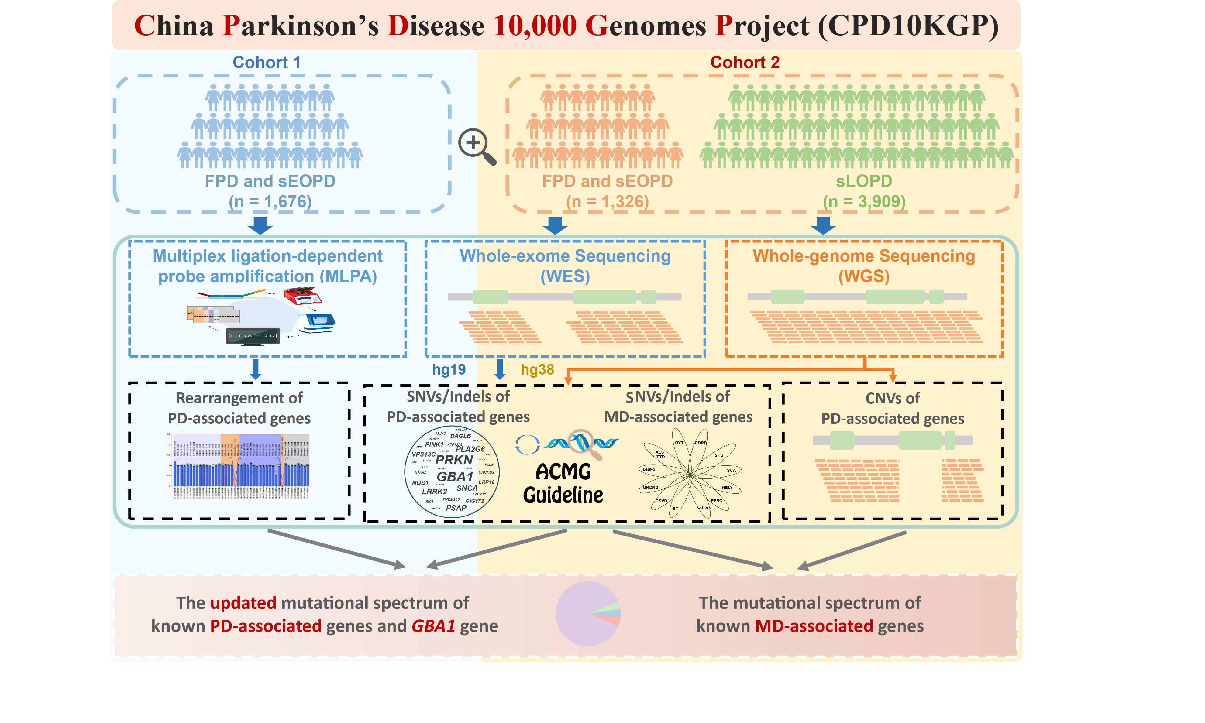
Fig 1. Research workflow of the present study.
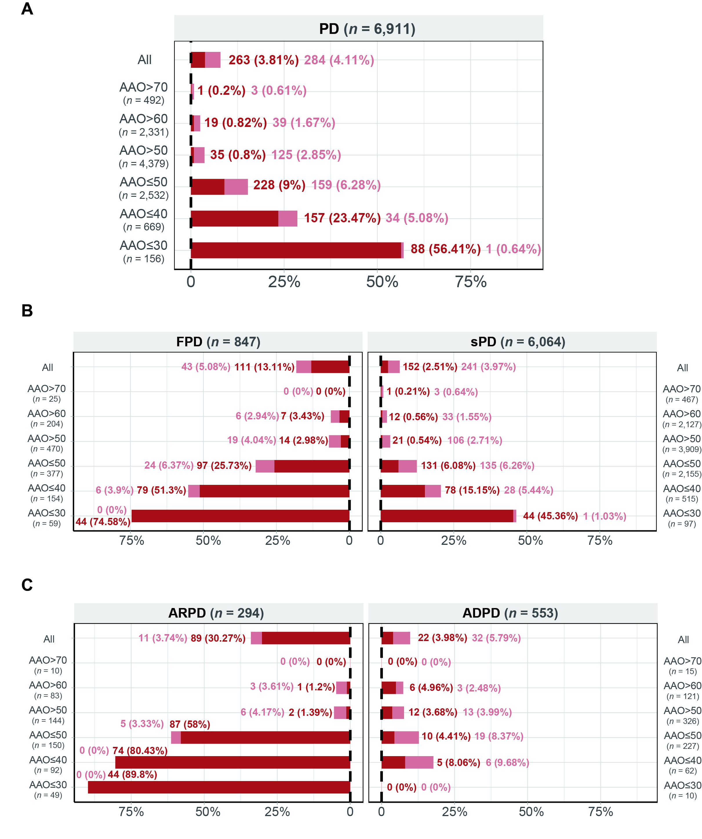
Fig 2. Mutational frequencies of each gene
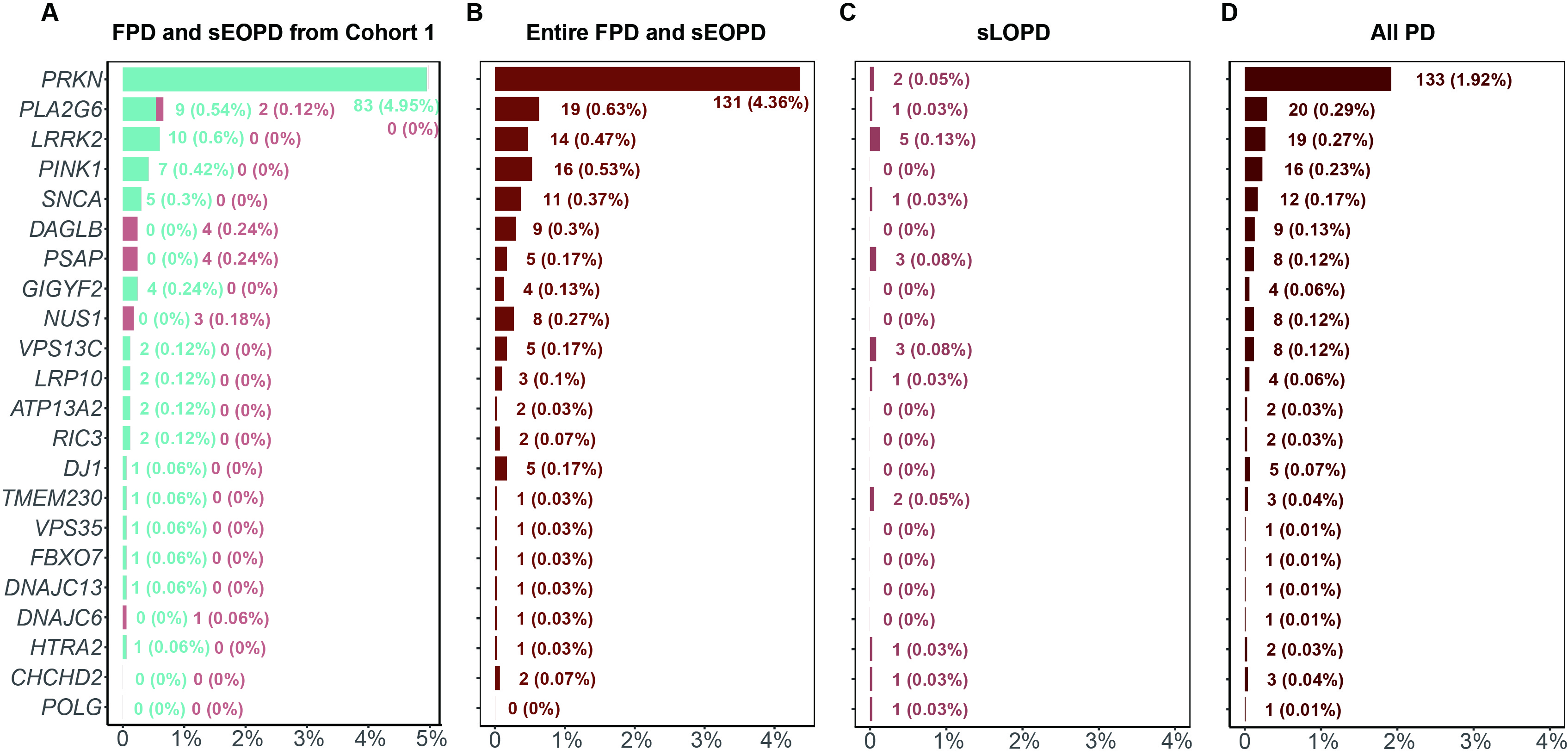
Fig 3. Mutational frequencies
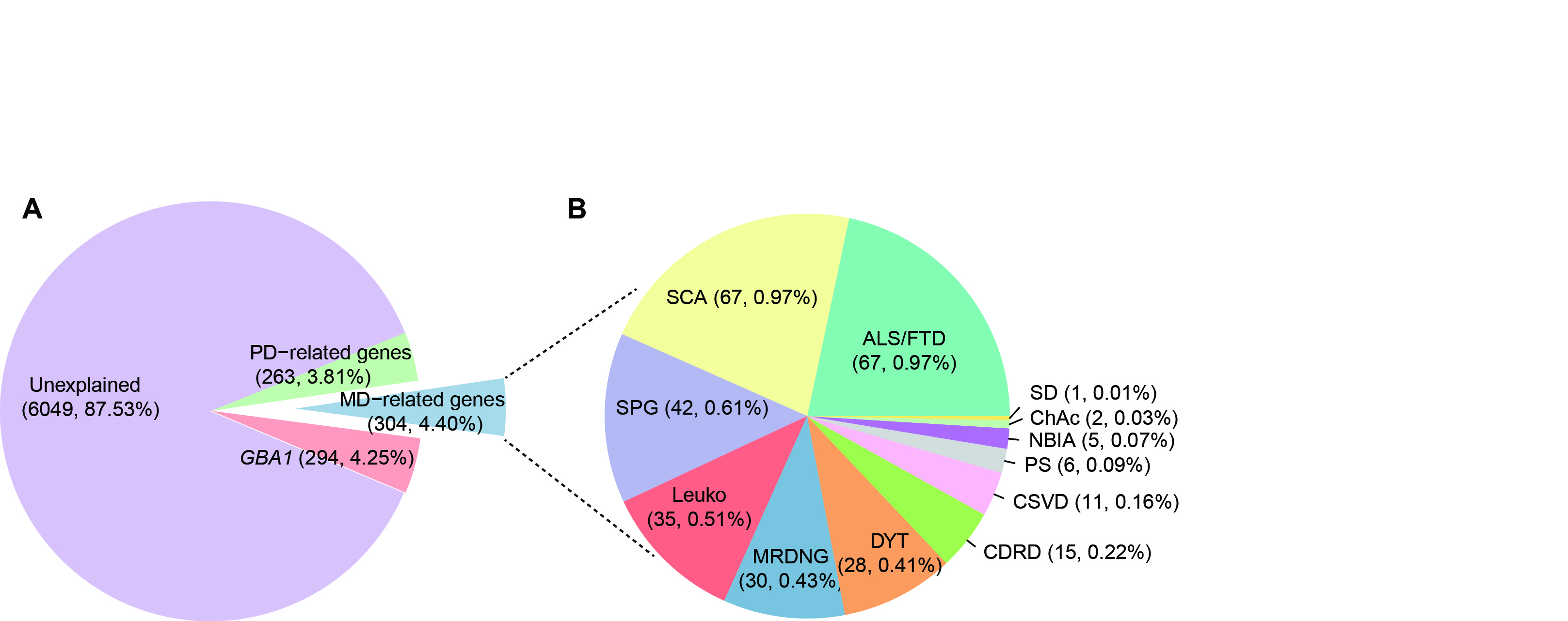
Fig 4. AAO spectrum
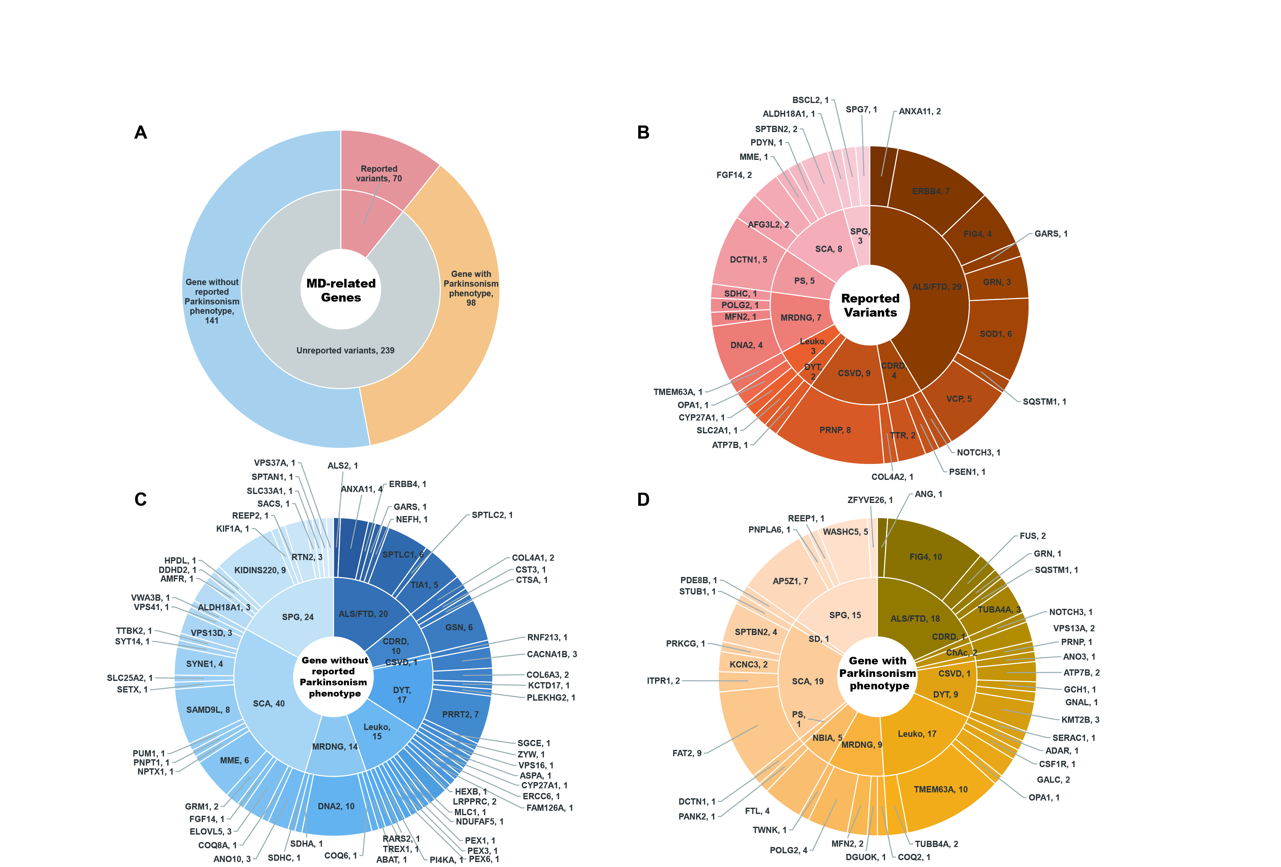
Fig 5. Circular visualization
References: 1. Tanner CM, Ostrem JL. Parkinson’s Disease. N Engl J Med. 2024; 391 (5): 442-452.
2. Morris HR, Spillantini MG, Sue CM, Williams-Gray CH. The pathogenesis of Parkinson’s disease. Lancet. 2024; 403 (10423): 293-304.
3. Lim SY, Tan AH, Ahmad-Annuar A, et al. Uncovering the genetic basis of Parkinson’s disease globally: from discoveries to the clinic. Lancet Neurol. 2024.
4. Pan H, Liu Z, Ma J, et al. Genome-wide association study using whole-genome sequencing identifies risk loci for Parkinson’s disease in Chinese population. NPJ Parkinsons Dis. 2023; 9 (1): 22.
5. Blauwendraat C, Nalls MA, Singleton AB. The genetic architecture of Parkinson’s disease. Lancet Neurol. 2020; 19 (2): 170-178.
6. Martin S, Smolders S, Van den Haute C, et al. Mutated ATP10B increases Parkinson’s disease risk by compromising lysosomal glucosylceramide export. Acta Neuropathol. 2020; 139 (6): 1001-1024.
7. Liu Z, Yang N, Dong J, et al. Deficiency in endocannabinoid synthase DAGLB contributes to early onset Parkinsonism and murine nigral dopaminergic neuron dysfunction. Nat Commun. 2022; 13 (1): 3490.
8. Hop PJ, Lai D, Keagle PJ, et al. Systematic rare variant analyses identify RAB32 as a susceptibility gene for familial Parkinson’s disease. Nat Genet. 2024; 56 (7): 1371-1376.
9. Gustavsson EK, Follett J, Trinh J, et al. RAB32 Ser71Arg in autosomal dominant Parkinson’s disease: linkage, association, and functional analyses. Lancet Neurol. 2024; 23 (6): 603-614.
10. Kara E, Tucci A, Manzoni C, et al. Genetic and phenotypic characterization of complex hereditary spastic paraplegia. Brain. 2016; 139 (Pt 7): 1904-1918.
11. Baizabal-Carvallo JF, Jankovic J. Parkinsonism, movement disorders and genetics in frontotemporal dementia. Nat Rev Neurol. 2016; 12 (3): 175-185.
12. Zhou X, Liu Z, Zhou X, et al. The Chinese Parkinson’s Disease Registry (CPDR): Study Design and Baseline Patient Characteristics. Mov Disord. 2022; 37 (7): 1335-1345.
13. Morris HR. Making neurogenetics a global endeavour. Brain. 2020; 143 (7): 1970-1973.
14. Zhao YW, Pan HX, Zeng Q, et al. PSAP variants in Parkinson’s disease: a large cohort study in Chinese mainland population. Brain. 2021; 144 (3): e25.
15. Zhao YW, Pan HX, Wang CY, et al. UQCRC1 variants in Parkinson’s disease: a large cohort study in Chinese mainland population. Brain. 2021; 144 (6): e54.
16. Zhao Y, Qin L, Pan H, et al. The role of genetics in Parkinson’s disease: a large cohort study in Chinese mainland population. Brain. 2020; 143 (7): 2220-2234.
17. Zhao Y, Pan H, Wang Y, et al. ATP10B variants in Parkinson’s disease: a large cohort study in Chinese mainland population. Acta Neuropathol. 2021; 141 (5): 805-806.
18. Richards S, Aziz N, Bale S, et al. Standards and guidelines for the interpretation of sequence variants: a joint consensus recommendation of the American College of Medical Genetics and Genomics and the Association for Molecular Pathology. Genet Med. 2015; 17 (5): 405-424.
19. Postuma RB, Berg D, Stern M, et al. MDS clinical diagnostic criteria for Parkinson’s disease. Mov Disord. 2015; 30 (12): 1591-1601.
20. Gibb WR, Lees AJ. The relevance of the Lewy body to the pathogenesis of idiopathic Parkinson’s disease. J Neurol Neurosurg Psychiatry. 1988; 51 (6): 745-752.
21. Li H. Toward better understanding of artifacts in variant calling from high-coverage samples. Bioinformatics. 2014; 30 (20): 2843-2851.
22. McKenna A, Hanna M, Banks E, et al. The Genome Analysis Toolkit: a MapReduce framework for analyzing next-generation DNA sequencing data. Genome Res. 2010; 20 (9): 1297-1303.
23. Yang H, Wang K. Genomic variant annotation and prioritization with ANNOVAR and wANNOVAR. Nat Protoc. 2015; 10 (10): 1556-1566.
24. Wang K, Li M, Hakonarson H. ANNOVAR: functional annotation of genetic variants from high-throughput sequencing data. Nucleic Acids Res. 2010; 38 (16): e164.
25. Wang Z, Zhao G, Zhu Z, et al. VarCards2: an integrated genetic and clinical database for ACMG-AMP variant-interpretation guidelines in the human whole genome. Nucleic Acids Res. 2024; 52 (D1): D1478-D1489.
26. Li J, Shi L, Zhang K, et al. VarCards: an integrated genetic and clinical database for coding variants in the human genome. Nucleic Acids Res. 2018; 46 (D1): D1039-D1048.
27. Ng PC, Henikoff S. SIFT: Predicting amino acid changes that affect protein function. Nucleic Acids Res. 2003; 31 (13): 3812-3814.
28. Adzhubei IA, Schmidt S, Peshkin L, et al. A method and server for predicting damaging missense mutations. Nat Methods. 2010; 7 (4): 248-249.
29. Chun S, Fay JC. Identification of deleterious mutations within three human genomes. Genome Res. 2009; 19 (9): 1553-1561.
30. Schwarz JM, Rodelsperger C, Schuelke M, Seelow D. MutationTaster evaluates disease-causing potential of sequence alterations. Nat Methods. 2010; 7 (8): 575-576.
31. Reva B, Antipin Y, Sander C. Predicting the functional impact of protein mutations: application to cancer genomics. Nucleic Acids Res. 2011; 39 (17): e118.
32. Kircher M, Witten DM, Jain P, O’Roak BJ, Cooper GM, Shendure J. A general framework for estimating the relative pathogenicity of human genetic variants. Nat Genet. 2014; 46 (3): 310-315.
33. Li J, Zhao T, Zhang Y, et al. Performance evaluation of pathogenicity-computation methods for missense variants. Nucleic Acids Res. 2018; 46 (15): 7793-7804.
34. Suvakov M, Panda A, Diesh C, Holmes I, Abyzov A. CNVpytor: a tool for copy number variation detection and analysis from read depth and allele imbalance in whole-genome sequencing. Gigascience. 2021; 10 (11).
35. Abyzov A, Urban AE, Snyder M, Gerstein M. CNVnator: an approach to discover, genotype, and characterize typical and atypical CNVs from family and population genome sequencing. Genome Res. 2011; 21 (6): 974-984.
36. DePristo MA, Banks E, Poplin R, et al. A framework for variation discovery and genotyping using next-generation DNA sequencing data. Nat Genet. 2011; 43 (5): 491-498.
37. Rausch T, Zichner T, Schlattl A, Stutz AM, Benes V, Korbel JO. DELLY: structural variant discovery by integrated paired-end and split-read analysis. Bioinformatics. 2012; 28 (18): i333-i339.
38. Layer RM, Chiang C, Quinlan AR, Hall IM. LUMPY: a probabilistic framework for structural variant discovery. Genome Biol. 2014; 15 (6): R84.
39. Jeffares DC, Jolly C, Hoti M, et al. Transient structural variations have strong effects on quantitative traits and reproductive isolation in fission yeast. Nat Commun. 2017; 8: 14061.
40. Geoffroy V, Herenger Y, Kress A, et al. AnnotSV: an integrated tool for structural variations annotation. Bioinformatics. 2018; 34 (20): 3572-3574.
41. Amberger JS, Hamosh A. Searching Online Mendelian Inheritance in Man (OMIM): A Knowledgebase of Human Genes and Genetic Phenotypes. Curr Protoc Bioinformatics. 2017; 58: 1 2 1-1 2 12.
42. Wang Y, Zhao Y, Pan H, et al. Genetic analysis of dystonia-related genes in Parkinson’s disease. Front Aging Neurosci. 2023; 15: 1207114.
43. Pan HX, Zhao YW, Mei JP, et al. GCH1 variants contribute to the risk and earlier age-at-onset of Parkinson’s disease: a two-cohort case-control study. Transl Neurodegener. 2020; 9 (1): 31.
44. Zeng Q, Pan H, Zhao Y, et al. Evaluation of common and rare variants of Alzheimer’s disease-causal genes in Parkinson’s disease. Parkinsonism Relat Disord. 2022; 97: 8-14.
45. Zeng Q, Pan H, Zhao Y, et al. Association between NOTCH3 gene and Parkinson’s disease based on whole-exome sequencing. Front Aging Neurosci. 2022; 14: 995330.
46. Liang D, Zhao Y, Pan H, et al. Rare variant analysis of essential tremor-associated genes in early-onset Parkinson’s disease. Ann Clin Transl Neurol. 2021; 8 (1): 119-125.
47. Zeng S, Zhou X, He R, et al. Association Analysis of Essential Tremor-Associated Genetic Variants in Sporadic Late-Onset Parkinson’s Disease. Tremor Other Hyperkinet Mov (N Y). 2024; 14: 25.
48. Zhou Y, Wang Y, Wan J, et al. Mutational spectrum and clinical features of GBA1 variants in a Chinese cohort with Parkinson’s disease. NPJ Parkinsons Dis. 2023; 9 (1): 129.
49. Guo JF, Zhang L, Li K, et al. Coding mutations in NUS1 contribute to Parkinson’s disease. Proc Natl Acad Sci U S A. 2018; 115 (45): 11567-11572.
50. Zhao Y, Pan H, Guo J, Tang B, Liu Z. RAB32 mutation in Parkinson’s disease. Lancet Neurol. 2024; 23 (10): 962-963.
51. Towns C, Fang ZH, Tan MMX, et al. Parkinson’s families project: a UK-wide study of early onset and familial Parkinson’s disease. NPJ Parkinsons Dis. 2024; 10 (1): 188.
52. Regensburger M, Turk M, Pagenstecher A, Schroder R, Winkler J. VCP-related multisystem proteinopathy presenting as early-onset Parkinson disease. Neurology. 2017; 89 (7): 746-748.
53. Kacem I, Funalot B, Torny F, Lautrette G, Andersen PM, Couratier P. Early onset Parkinsonism associated with an intronic SOD1 mutation. Amyotroph Lateral Scler. 2012; 13 (3): 315-317.
54. Esteves T, Durr A, Mundwiller E, et al. Loss of association of REEP2 with membranes leads to hereditary spastic paraplegia. Am J Hum Genet. 2014; 94 (2): 268-277.
55. Nudelman KNH, Jackson T, Rumbaugh M, et al. Pathogenic variants in the Longitudinal Early-onset Alzheimer’s Disease Study cohort. Alzheimers Dement. 2023; 19 Suppl 9 (Suppl 9): S64-S73.
56. Westenberger A, Skrahina V, Usnich T, et al. Relevance of genetic testing in the gene-targeted trial era: the Rostock Parkinson’s disease study. Brain. 2024; 147 (8): 2652-2667.
57. Cook L, Verbrugge J, Schwantes-An TH, et al. Parkinson’s disease variant detection and disclosure: PD GENEration, a North American study. Brain. 2024; 147 (8): 2668-2679.
58. Verdura E, Rodriguez-Palmero A, Velez-Santamaria V, et al. Biallelic PI4KA variants cause a novel neurodevelopmental syndrome with hypomyelinating leukodystrophy. Brain. 2021; 144 (9): 2659-2669.
59. Parmar JM, McNamara EL, Lamont PJ, et al. Two Novel Variants in PI4KA in a Family Presenting With Hereditary Spastic Paraparesis: A Case Report. Neurol Genet. 2024; 10 (3): e200152.
60. Tsoi H, Yu AC, Chen ZS, et al. A novel missense mutation in CCDC88C activates the JNK pathway and causes a dominant form of spinocerebellar ataxia. J Med Genet. 2014; 51 (9): 590-595.
61. Caputo D, Cetica V, Paoli S, Rosati A, Lazzeri S. Confirmation of the Pathogenetic Role of the CCDC88C Gene in Early-Onset Pure Spastic Paraplegia. Mov Disord. 2023; 38 (8): 1561-1562.
62. Tappino B, Biancheri R, Mort M, et al. Identification and characterization of 15 novel GALC gene mutations causing Krabbe disease. Hum Mutat. 2010; 31 (12): E1894-1914.
63. Milovanovic A, Westenberger A, Stankovic I, et al. ANO10-Related Spinocerebellar Ataxia: MDSGene Systematic Literature Review and a Romani Case Series. Mov Disord. 2024; 39 (5): 887-892.
To cite this abstract in AMA style:
Y. Zhao, Z. Liu, H. Pan, J. Guo, B. Tang. Unveiling Parkinson’s Disease Variants in the Chinese Population: The CPD10KGP Study [abstract]. Mov Disord. 2025; 40 (suppl 1). https://www.mdsabstracts.org/abstract/unveiling-parkinsons-disease-variants-in-the-chinese-population-the-cpd10kgp-study/. Accessed April 10, 2026.« Back to 2025 International Congress
MDS Abstracts - https://www.mdsabstracts.org/abstract/unveiling-parkinsons-disease-variants-in-the-chinese-population-the-cpd10kgp-study/