Session Information
Date: Wednesday, September 25, 2019
Session Title: Neuroimaging
Session Time: 1:15pm-2:45pm
Location: Les Muses Terrace, Level 3
Objective: To study the anatomic volume differences of essential tremor (ET) patient before and after MR-guided focused ultrasound (MRgFUS) thalamotomy using respective 3D T1-weighted MR image.
Background: ET is one of the most common neurological disorders, and it might affect the hand, leg, trunk, or vocal cord with an involutory tremor to the patient. Medical refractory ET can be effectively controlled by MRgFUS thalamotomy, which causes the thermo-coagulation at the contralateral side of the ventral intermediate nucleus (VIM) of the thalamus to the treated hand. MRgFUS thalamotomy caused the functional changed in the brain, but no literature revealed the anatomic changed after receiving this treatment.
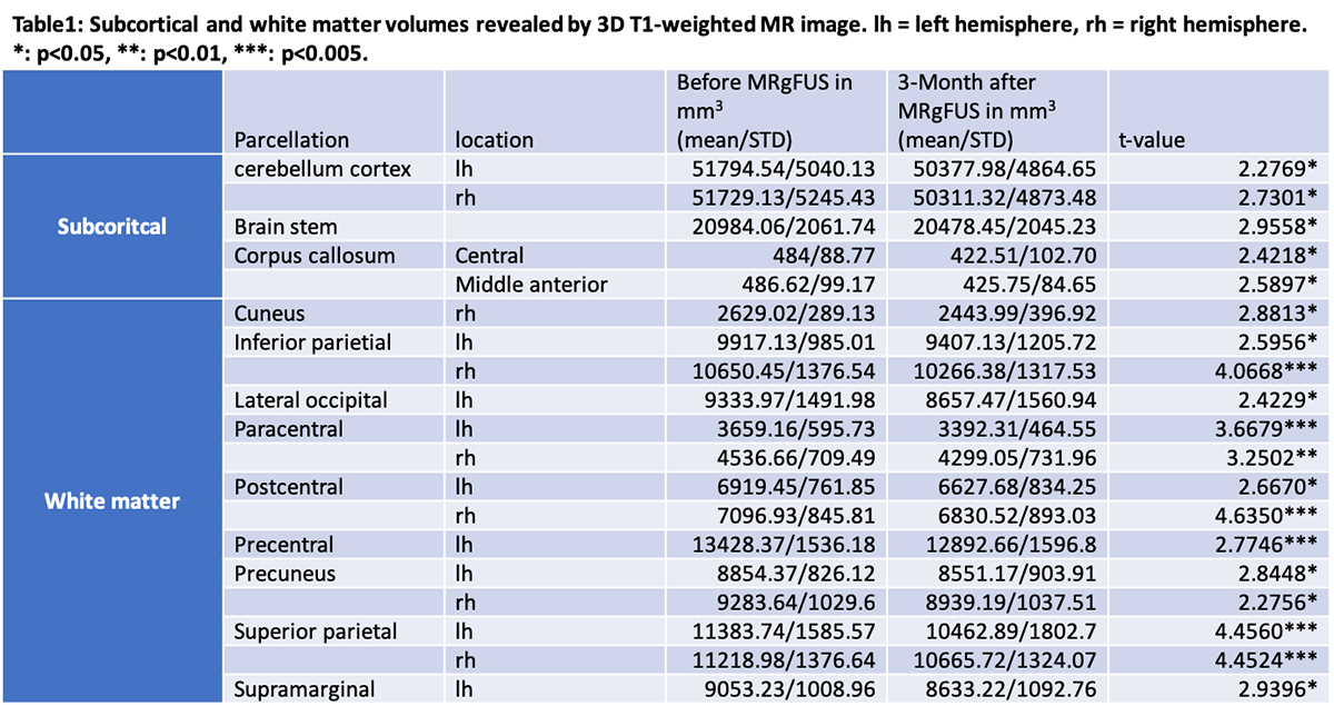
Method: Thirteen ET patients (1 female and 12 male, mean/STD of age=58/12.2 years old) received 1 mm isotropic 3D T1-weighted MR imaging (BRAVO) with a 1.5T MR scanner (GE, USA) one day before and three months after the left VIM MRgFUS (Insightec, Israel) thalamotomy to mitigate their right-hand tremors. The FreeSurfer was used to reconstruct each volumetric MR image and estimate the volume in mm3of the subcortical and white matter at the different anatomic parcellations. Two-tailed paired t-test was used to exam the difference between anatomic parcellations before and after the MRgFUS thalamotomy across all patients.
Results: In subcortical parcellations, the bilateral cerebellum cortex, brain stem, and central and middle anterior part of corpus callosum showed significant volume decrements after receiving MRgFUS thalamotomy. In white matter parcellations, the bilateral inferior parietal, paracentral, postcentral, precuneus, superior parietal, as well as right-side cuneus, and left-side lateral occipital, precentral, and supramarginal showed significant volume decrements after receiving MRgFUS thalamotomy. Table1summarized the mean and STD volumes, as well as the statistics of all the parcellations.
Conclusion: In this study we found several parcellation volumes in the brain decreased after receiving MRgFUS thalamotomy. This anatomic volume analysis provides another figure of merit to estimate the long-term effect of novel MRgFUS thalamotomy to control ET.
To cite this abstract in AMA style:
K. Tsai, HC. Lai, CY. Wei, PY. Chiu, W. Lin, CL. Chen, SK. Yang, CH. Hung, WC. Chang. White matter and subcortical volume of essential tremor patient decreases after MR-guided focused ultrasound thalamotomy [abstract]. Mov Disord. 2019; 34 (suppl 2). https://www.mdsabstracts.org/abstract/white-matter-and-subcortical-volume-of-essential-tremor-patient-decreases-after-mr-guided-focused-ultrasound-thalamotomy/. Accessed March 26, 2026.« Back to 2019 International Congress
MDS Abstracts - https://www.mdsabstracts.org/abstract/white-matter-and-subcortical-volume-of-essential-tremor-patient-decreases-after-mr-guided-focused-ultrasound-thalamotomy/