Category: Ataxia
Objective: To assess and quantify microstructural white matter atrophy in brain of spinocerebellar ataxia (SCA) types SCA1 and SCA2 patients in comparison with healthy subjects.
Background: White matter pathways, including the cortico-ponto-cerebello-thalamo-cortical and cortico-cerebello-cortical loops, and cerebellum are affected in autosomal dominant SCAs. These pathways play a crucial role in cognitive and motor function [1]. Mid brain, brainstem and cerebellum atrophies are reported in SCA1 and SCA2 patients using MR imaging [2]. Clinical neuroradiologic differentiation of SCA1 and SCA2 are very difficult [3], as disease progression is fast in SCA1 and 2 than other SCAs, resulting in increased white matter volume loss in brain stem and cerebellum [4].
We used diffusion tensor imaging (DTI) to estimate white matter tract volume in 42 tracts (19 left/ 19 right, Forceps Major, Forceps Minor, Middle Cerebellar Peduncle, and Anterior Commissure) based on probabilistic tract atlas.
Method: Symptomatic and genetically confirmed SCA1 (n= 27), SCA2 (n=25) patients assessed with International Cooperative Ataxia Rating Scale (ICARS,) and healthy controls (HC, n=28) were recruited (Table 1).
DTI data was acquired on a 3T MR scanner (Ingenia 3.0 T, M/s. Philips Healthcare, The Netherlands) using 32-channel head coil, single-shot echo-planar dual SE sequence in 32 directions, b= 0 and 800 s/mm2, slice thickness=2 mm with no gap, number of slices=74, FOV=230 mm, TR = 4802 ms, EPI factor 39, echo spacing: 0.69, TE:92ms, averages:2, Flip angle: 90◦, Phase encoding direction: A-P and Bandwidth: 1860Hz.
Results: FA values were decreased in bilateral corticospinal tract, Forceps Minor, Middle Cerebellar Peduncle, right Optic Radiation, left Uncinate Fasciculus, and Anterior Thalamic Radiation in SCA1 and SCA2 groups in comparison with HC (Tables 2 and Figures 1). WM tract volume is reduced in Forceps Major in SCA1, and left Cingulum subsection Dorsal and right Frontal Aslant in SCA2 in comparision with HC. We observed increased RD and AD values in bilateral anterior thalamic radiation and corticospinal tracts in SCA1 and SCA2 as compared to HC.
Conclusion: Decreased white matter volume and FA values in corticospinal and thalamic tracts (with respect to that in healthy controls) may be associated with increased motor dysfunction [5,6,7], cognitive and behavioural impairments in SCA1 and SCA2 [8].
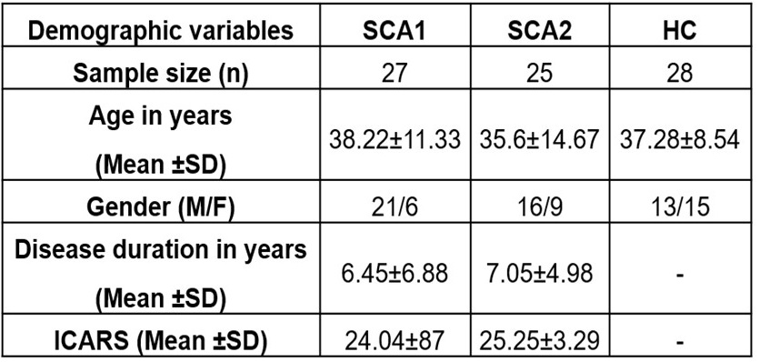
Demographic details in SCA1, SCA2 and HC.
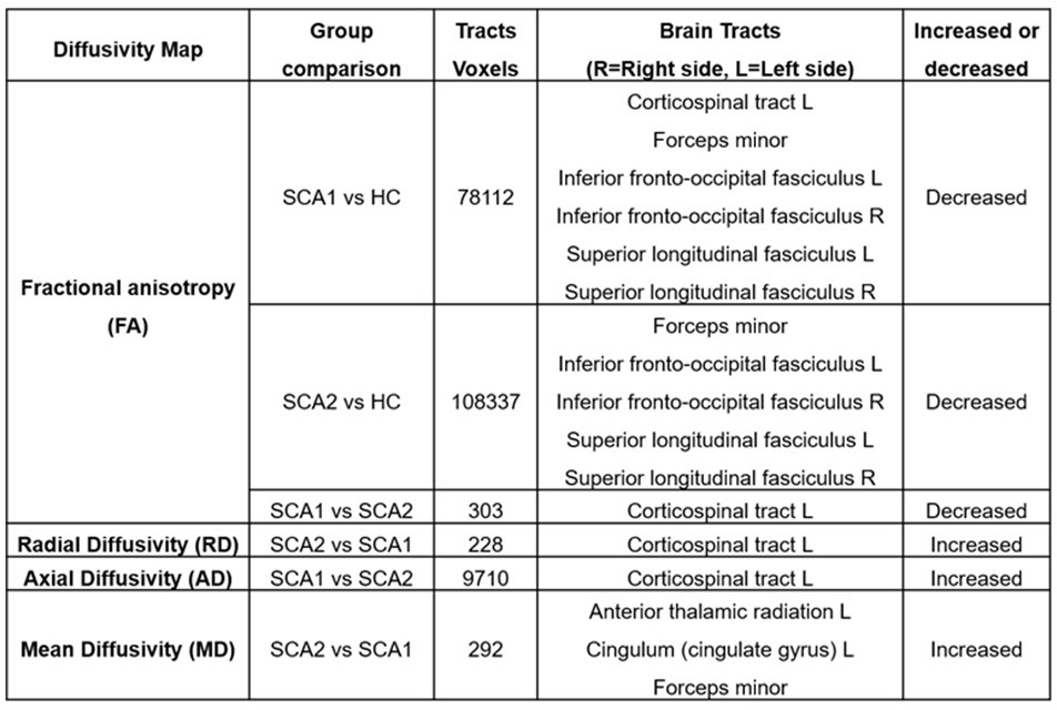
Changes in diffusivity parameters in SCA and HC.
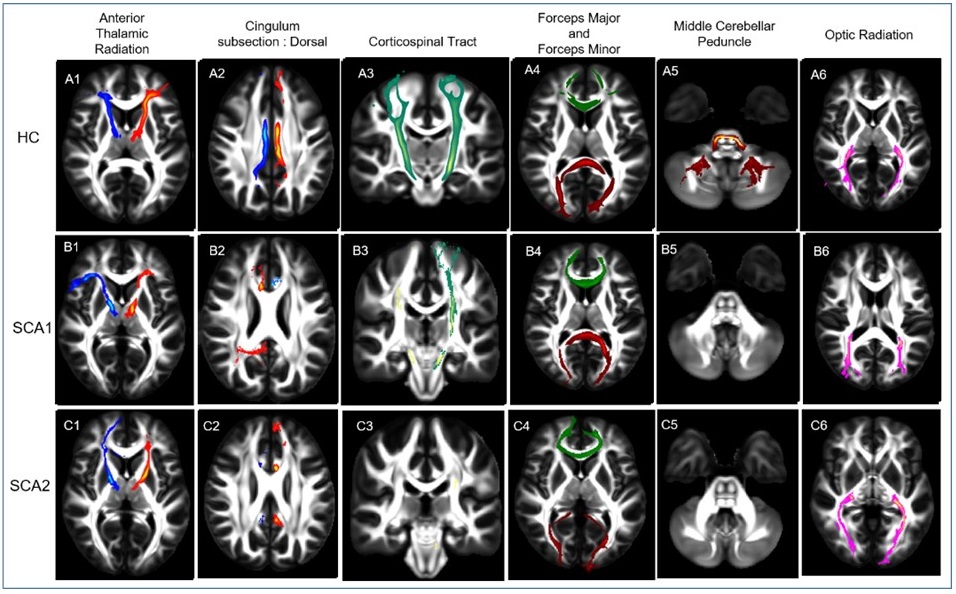
DTI based tractography of white matter SCA and HC.
References: 1. Palesi F, Tournier JD, Calamante F, et al. Contralateral cerebello-thalamo-cortical pathways with prominent involvement of associative areas in humans in vivo. Brain Struct Funct. 2015 Nov;220(6):3369-84.
2. Della Nave R, Ginestroni A, Tessa C, et al. Brain white matter damage in SCA1 and SCA2. An in vivo study using voxel-based morphometry, histogram analysis of mean diffusivity and tract-based spatial statistics. Neuroimage. 2008 Oct 15;43(1):10-9.
3. Manto, M. and Habas, C., 2016. Cerebellar disorders: clinical/radiologic findings and modern imaging tools. Handbook of Clinical Neurology, 135, pp.479-491.
4. Martins Junior CR, Martinez ARM, Vasconcelos IF, et al. Structural signature in SCA1: clinical correlates, determinants and natural history. J Neurol. 2018 Dec;265(12):2949-2959.
5. Morales H, Tomsick T. Middle cerebellar peduncles: Magnetic resonance imaging and pathophysiologic correlate. World J Radiol. 2015 Dec 28;7(12):438-47.
6. Meira AT, Arruda WO, Ono SE, et al. Neuroradiological Findings in the Spinocerebellar Ataxias. Tremor Other Hyperkinet Mov (N Y). 2019 Sep 26;9. doi: 10.7916/tohm.v0.682.
7. Alcauter S, Barrios FA, Díaz R, et al. Gray and white matter alterations in spinocerebellar ataxia type 7: an in vivo DTI and VBM study. Neuroimage. 2011 Mar 1;55(1):1-7.
8. Stezin A, Bhardwaj S, Khokhar S, et al. In vivo microstructural white matter changes in early spinocerebellar ataxia 2. Acta Neurol Scand. 2021 Mar;143(3):326-332.
To cite this abstract in AMA style:
P. Pankaj, A. Srivastava, M. Kumar, S. Kumaran, A. Garg, R. Agarwal, A. Nehra. White Matter Tractography in Spinocerebellar Ataxia type 1 and 2 in comparison with Healthy Controls [abstract]. Mov Disord. 2024; 39 (suppl 1). https://www.mdsabstracts.org/abstract/white-matter-tractography-in-spinocerebellar-ataxia-type-1-and-2-in-comparison-with-healthy-controls/. Accessed June 18, 2026.« Back to 2024 International Congress
MDS Abstracts - https://www.mdsabstracts.org/abstract/white-matter-tractography-in-spinocerebellar-ataxia-type-1-and-2-in-comparison-with-healthy-controls/