Session Information
Date: Monday, October 8, 2018
Session Title: Parkinson's Disease: Psychiatric Manifestations
Session Time: 1:15pm-2:45pm
Location: Hall 3FG
Objective: To investigate patterns of cerebral blood flow alteration associated with depressive state in Parkinson’s disease (PD) by using principal component analysis of arterial spin labelling (ASL) MRI as a non-invasive methodology of cerebral blood flow (CBF) mapping.
Background: Invasive nuclear imaging has demonstrated reduced CBF in discrete brain regions of PD with depression. PD affects a large range of neural-pathways and neurotransmitters and disease-related CBF patterns were successfully demonstrated using ASL and a multivariate pattern analysis technique. To our knowledge, this is the first study reporting ASL findings of perfusion patterns in PD with depression using principal component analysis (PCA).
Methods: 45 PD patients underwent whole-brain ASL and high-resolution T1-weighted 3D MRI scan at 3T (Discovery MR 750, GE Healthcare, US). CBF maps were obtained and realigned to a standard template and then normalized and smoothed. The Beck Depression Inventory (BDI-II) score was used to group participants into non-or-minimal depressed PD (BDI-II <13) [n=30, mean age: 68.6±7.4; 22 male] and mild to severe depression (BDI-II >13) [n=15, mean age: 65±7.1; 9 male]. The resulting CBF maps were investigated by voxel-based PCA to identify group differential CBF patterns.
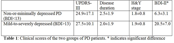
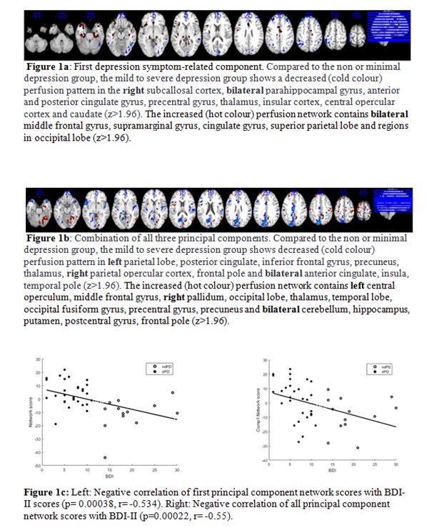
Results: Subgroups did not differ in demographic, PD disease duration or motor severity (Table 1). Absolute CBF of global grey matter (including cerebellum) was non-significantly lower in depressed (40.5±7.5ml/100g/min) vs. non-depressed subgroups (43.3±6.3ml/100g/min, p=0.192). PCA revealed a distinct perfusion pattern that differentiated both groups, a linear combination of three components (area under the curve= 0.86). The network scores were negatively correlated with individual BDIs by controlling for sex and age (Figure 1). [table1] [figure1]
Conclusions: Reduced activity is shown in the limbic and cortico-basal ganglia-thalamic networks of the mild-severe depression vs. the non-or-minimal depression group. This is partially in line with previous PET studies, but also suggests that different components might allow differentiation of subtypes of depressive states. In conclusion, our study suggests that combining non-invasive CBF mapping using ASL with PCA allows identification of patterns of network dysfunction that relate to non-motor symptoms in patients with PD.
References: Melzer, T. R. et al. (2011) ‘Arterial spin labelling reveals an abnormal cerebral perfusion pattern in Parkinson’s disease. Brain. doi: 10.1093/brain/awq377.
To cite this abstract in AMA style:
Y. Xing, S. Naidu, S. Schwarz, N. Bajaj, D. Auer. Brain Perfusion pattern linked to depressive state in Parkinson’s: An arterial spin labelling MRI study [abstract]. Mov Disord. 2018; 33 (suppl 2). https://www.mdsabstracts.org/abstract/brain-perfusion-pattern-linked-to-depressive-state-in-parkinsons-an-arterial-spin-labelling-mri-study/. Accessed June 23, 2026.« Back to 2018 International Congress
MDS Abstracts - https://www.mdsabstracts.org/abstract/brain-perfusion-pattern-linked-to-depressive-state-in-parkinsons-an-arterial-spin-labelling-mri-study/