Category: Parkinsonism (Other)
Objective: To describe the clinical features, diagnostic outcomes, and brain MRI imaging characteristics in patients with ventriculomegaly and gait impairment
Background: Emerging evidence suggests that ventriculomegaly, commonly seen in idiopathic normal pressure hydrocephalus (iNPH), also occurs in neurodegenerative diseases like Alzheimer’s disease (AD), Parkinson’s disease (PD), dementia with Lewy bodies (DLB), and progressive supranuclear palsy (PSP). Imaging measures such as Evans’s index (EI), Callosal angle (CA), and Disproportionately Enlarged Subarachnoid Space Hydrocephalus (DESH) are helpful, but differences in neurodegenerative contexts remain unclear
Method: A retrospective study analyzed 55 patients with gait impairment and 40 matched sex/age healthy controls (HCs). Brain MRIs assessed ventriculomegaly using EI>0.3 and CA<90º, alongside DESH. Patients were classified into neurodegeneration (PSP, AD, DLB, PD), iNPH, and HCs based on diagnostic criteria, cognitive tests, CSF biomarkers, and ¹⁸F-Dopa-PET during follow-up. Kruskal-Wallis and Bonferroni tests were used for analysis
Results: Clinical features and follow-up assessments, including ancillary tests (cognitive evaluations, biomarkers, and PET imaging), are summarized in [Tables 1-4]. The CA was lower in neurodegeneration (ND) patients compared to iNPH and HCs (79.9 ± 5.2 vs 87.5 ± 2.0 vs 129.7 ± 3.6, p <0.001). EI was similar between ND and iNPH groups. The DESH was higher in ND patients compared to iNPH and HCs (6.5 ± 1.3 vs 4.5 ± 0.9 vs 0.10 ± 0.3, p <0.001) [Table 5]
Conclusion: Our findings suggest that ventriculomegaly, coupled with neurodegeneration, is associated with lower CA and higher DESH, reinforcing the potential role of ventriculomegaly in neurodegenerative disorders, though caution is needed for shunting decisions
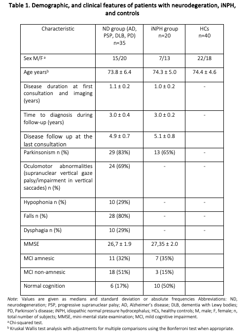
Demographic, and clinical features of patients
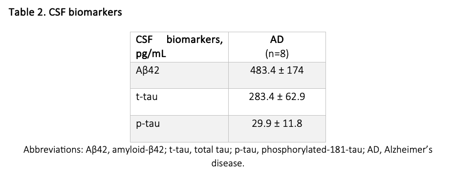
CSF biomarkers
![[18F]-dopa PET and L-dopa response](https://www.mdsabstracts.org/wp-content/uploads/2025/09/0936_1402_000796_3.png)
[18F]-dopa PET and L-dopa response
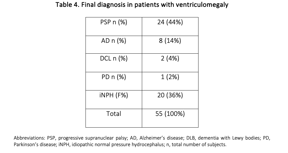
Final diagnosis in patients with ventriculomegaly
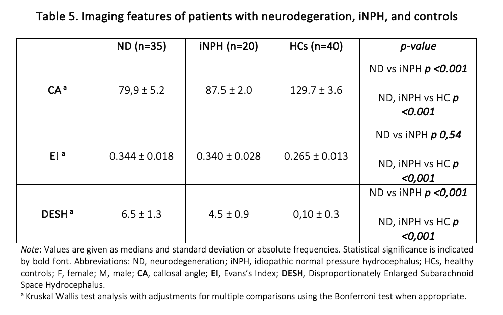
Imaging features of patients
References: 1. Espay AJ, Da Prat GA, Dwivedi AK, Rodriguez-Porcel F, Vaughan JE, Rosso M, Devoto JL, Duker AP, Masellis M, Smith CD, Mandybur GT, Merola A, Lang AE. Deconstructing normal pressure hydrocephalus: Ventriculomegaly as early sign of neurodegeneration. Ann Neurol. 2017 Oct;82(4):503-513. doi: 10.1002/ana.25046. Epub 2017 Oct 4. PMID: 28892572
2. Relkin N, Marmarou A, Klinge P, Bergsneider M, Black PM. Diagnosing idiopathic normal-pressure hydrocephalus. Neurosurgery. 2005 Sep;57(3 Suppl):S4-16; discussion ii-v. doi: 10.1227/01.neu.0000168185.29659.c5. PMID: 16160425
3. Fu MH, Huang CC, Wu KLH, Chen YF, Kung YC, Lee CC, Liu JS, Lan MY, Chang YY. Higher prevalence of idiopathic normal pressure hydrocephalus-like MRI features in progressive supranuclear palsy: An imaging reminder of atypical parkinsonism. Brain Behav. 2023 Feb;13(2):e2884. doi: 10.1002/brb3.2884. Epub 2023 Jan 12. PMID: 36635882; PMCID: PMC9927835.
4. Ohara M, Hattori T, Yokota T. Progressive supranuclear palsy often develops idiopathic normal pressure hydrocephalus-like magnetic resonance imaging features. Eur J Neurol. 2020 Oct;27(10):1930-1936. doi: 10.1111/ene.14322. Epub 2020 Jun 29. PMID: 32416639.
To cite this abstract in AMA style:
C. Espinoza-Vinces, I. Avilés-Olmos, J. Núñez Córdoba, M. Jiménez Vázquez, M. Calvo Imirizaldu, G. Martí-Andrés, M. Luquin. Analysis of Diagnostic Outcomes and Imaging Features in Patients with Ventriculomegaly and Gait Impairment [abstract]. Mov Disord. 2025; 40 (suppl 1). https://www.mdsabstracts.org/abstract/analysis-of-diagnostic-outcomes-and-imaging-features-in-patients-with-ventriculomegaly-and-gait-impairment/. Accessed April 6, 2026.« Back to 2025 International Congress
MDS Abstracts - https://www.mdsabstracts.org/abstract/analysis-of-diagnostic-outcomes-and-imaging-features-in-patients-with-ventriculomegaly-and-gait-impairment/