Category: Parkinson's Disease: Neuroimaging
Objective: Utilization of visual field testing and diffusion tensor imaging (DTI) to assess visual field deficits in patients with moderate-to-severe Parkinson’s Disease (PD) and identify biomarkers in the primary visual pathway controlling impairment.
Background: Visual field defects in PD patients are well-established, including color vision changes, retinal dopaminergic insufficiency, and visuospatial and visuoperceptual impairments [1], stemming from multi-level system dysfunction. Studies have further demonstrated the morphological changes associated with PD in the visual system such as retinal thinning [1], among others. Despite extensive establishment of visual field defects and neural damage seen in PD, limited studies have attempted to correlate damaged visual pathways in PD to the visual field impairments observed.
Method: A total of 5 PD patients 45+ diagnosed with moderate-to-severe PD were recruited in this study. Subjects were compared to age-matched controls without other neurological syndromes, pain disorders, cancer, or uncontrolled diabetes. DTI and visual field testing were performed on all subjects. Data analysis aims to determine average regional visual field degradation in each eye and correlate data with DTI tractography to identify hallmark changes in the primary visual pathway.
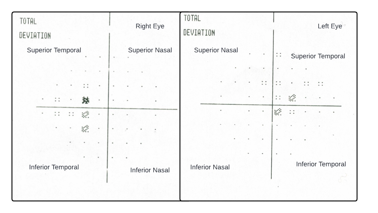
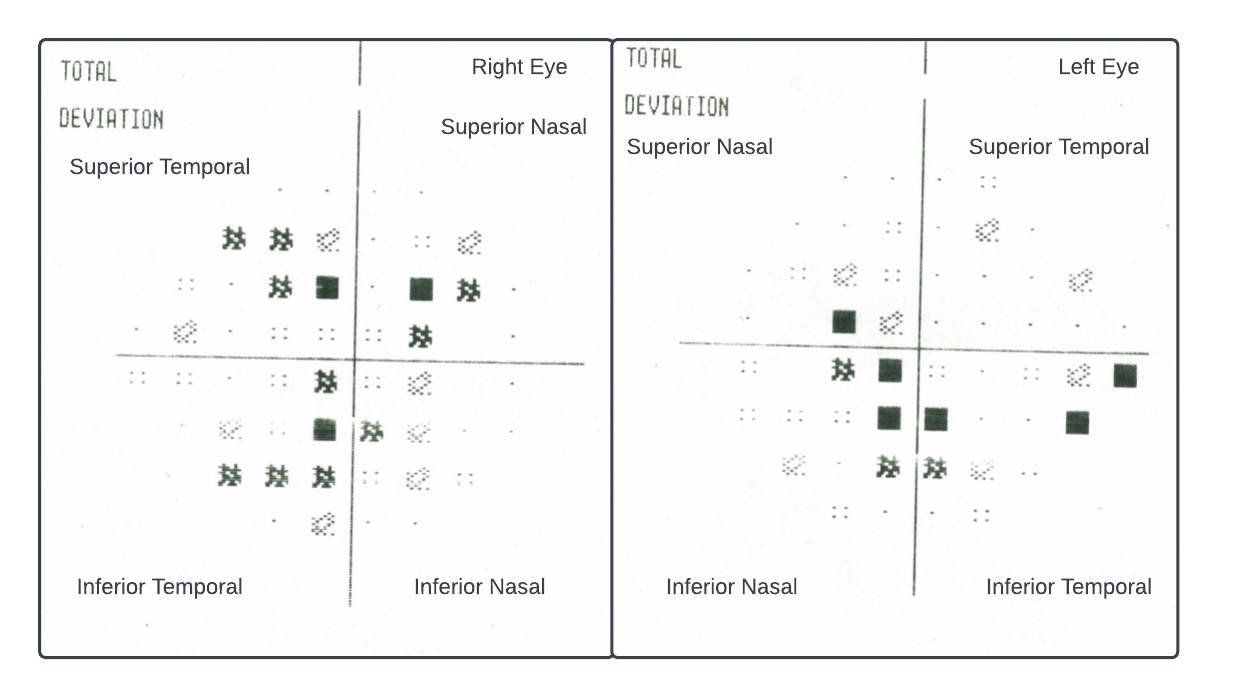
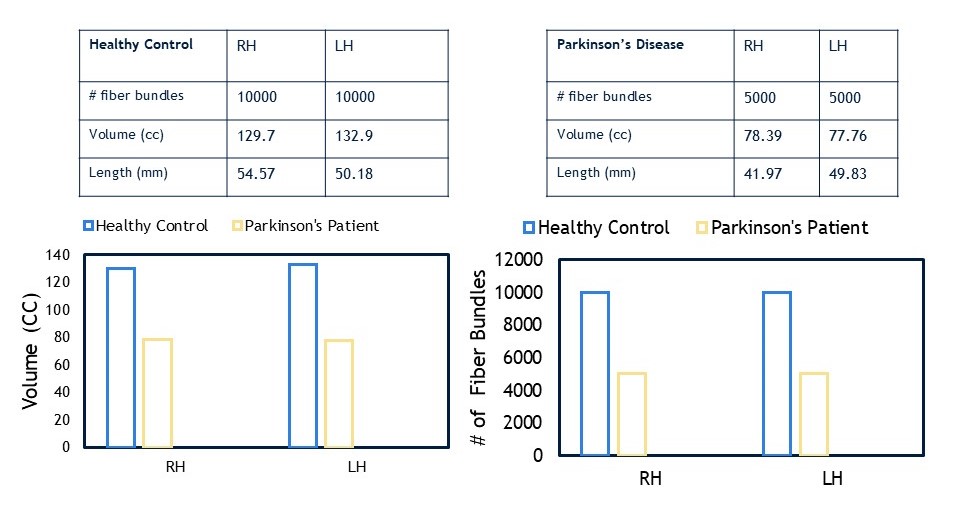
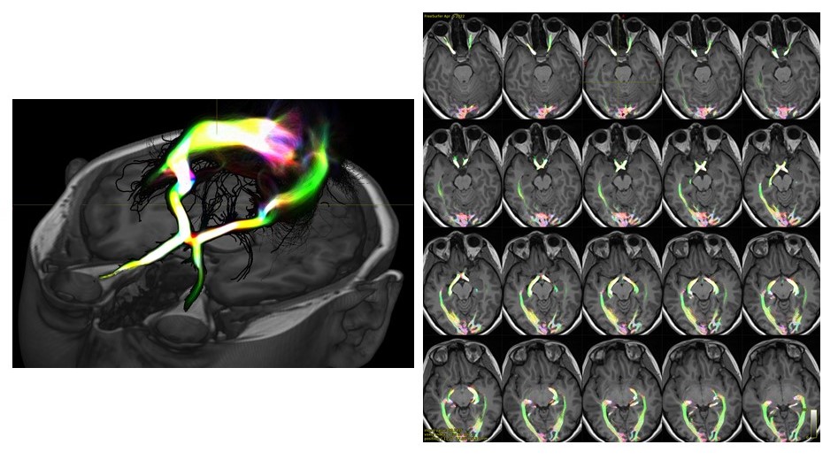
Results: Preliminary data collection has been performed for one control and five PD subjects. The healthy control showed low grade temporal field impairment in both eyes [Figure 1]. These deficits can be discerned by the shading in the diagram, with darker colors indicating severe impairment. General trends among PD patients unveil a greater degree of perceptual deficit in the temporal fields of both eyes, particularly the inferior portions. [Figure 2] portrays sample PD data. The primary visual pathway was reconstructed using probabilistic constrained spherical deconvolution (CSD) based diffusion tractography to identify demyelination and atrophy of primary visual pathway as well as decreases in fiber density and length [Figure 3]. [Figure 4] highlights an example DTI from a healthy control.
Conclusion: Current data implies that PD patients demonstrate accelerated perceptual losses in the temporal visual fields, particularly the inferior portions. Upon further data collection and analysis, we aim to extrapolate biomarkers from DTI data and unveil additional trends in visual field impairment.
Figure 1
Figure 2
Figure 3
Figure 4
References: 1. Ozlem Yenice, Sumru Onal, Ipek Midi, Eda Ozcan, Ahmet Temel, Dilek I-Gunal, Visual field analysis in patients with Parkinson’s disease, Parkinsonism & Related Disorders, Volume 14, Issue 3, 2008, Pages 193-198, ISSN 1353-8020, https://doi.org/10.1016/j.parkreldis.2007.07.018.
To cite this abstract in AMA style:
S. Mehta, M. Syed, S. Naghizadehkashani, K. Shivok, L. Krisa, C. Matias, C. Wu, T. Liang, R. Sergott, M. Alizadeh. Assessment of Visual Field in Patients with Moderate-to-Severe Parkinson’s Disease [abstract]. Mov Disord. 2024; 39 (suppl 1). https://www.mdsabstracts.org/abstract/assessment-of-visual-field-in-patients-with-moderate-to-severe-parkinsons-disease/. Accessed July 13, 2025.« Back to 2024 International Congress
MDS Abstracts - https://www.mdsabstracts.org/abstract/assessment-of-visual-field-in-patients-with-moderate-to-severe-parkinsons-disease/


