Session Information
Date: Tuesday, September 24, 2019
Session Title: Parkinsonisms and Parkinson-Plus
Session Time: 1:45pm-3:15pm
Location: Agora 3 West, Level 3
Objective: The aim of this study was to test using multimodal MRI biomarkers if the pattern of neurodegeneration in the brain of patients with PD fits the model of disease progression proposed by Braak and al. (2003).
Background: Parkinson’s disease (PD) is a progressive neurodegenerative illness characterized by extensive dopaminergic neurons damage in the substantia nigra (SN), accompanied by extensive extranigral pathology. Braak et al. (2003) proposed a staging model, based on neuropathology, suggesting that damage extended progressively in 6 stages from the medulla oblongata to cortical structures. This model has been challenged. There is also a heterogeneity of the disease with some patients presenting more severe forms of the disease, for instance presenting rapid eye movement sleep behavior disorder (RBD).
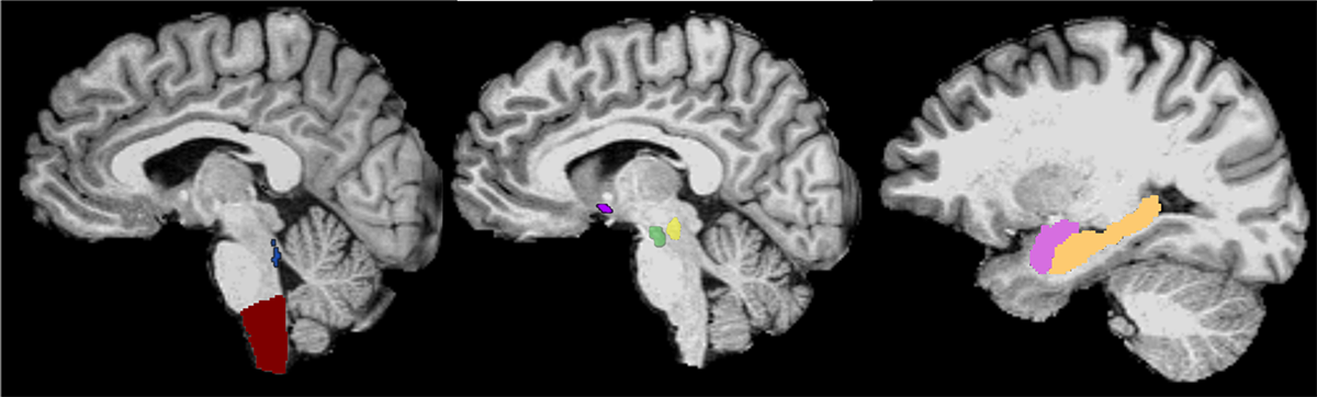
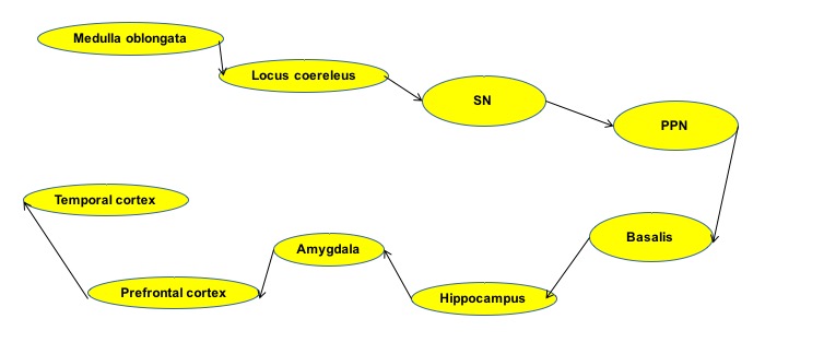
Method: 54 patients with PD, including 34 PD-RBD were compared with 25 healthy volunteers. MRI acquisition included 3D T1-w images, DTI and T1-w neuromelanin-sensitive (NM) images. The regions of interest (ROIs) were segmented in cortical and subcortical regions, in the SN, the basalis, the locus coeruleus (LC), and pedunculopontine nucleus (PPN) (Fig. 1). Volume, neuromelanin signal, mean diffusivity (MD), axial diffusivity (AD) and radial diffusivity (RD) were calculated in the ROIs. Statistical analysis: Partial Least Squares Path-Modeling (PLS-PM) was used to build and test an analytical path model based on Braak staging (Fig 2). PLS-PM is an approach to Structural Equation Modeling to estimate a network of causal relationships between blocks of variables. The overall quality of the model was assessed using Goodness of fit coefficient (Gof).
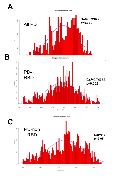
Results: Patients with PD showed increased MD, AD and LD in the medulla oblongata, PPN, basalis, hippocampus and amygdala. There was a significant decrease in LC-NM signal and SN-NM volume. PLS-PM was run first on all patients, then PD-RBD vs no RBD. The Gof was significant for all patient groups indicating agreement with Braak’s model (0.72, p=0.002). The PD-RBD group showed the highest Gof (0.74, p=0.002) while Gof in PD without RBD was not significant (0.7, p>0.05, Fig 3).
Conclusion: Using PLS-PM multi-modal imaging-based model, we found that the pattern of neurodegeneration in PD patients fitted Braak’s model of disease progression in PD patients with RBD (who probably had greater extent of damage in the brainstem) more than in patients without RBD.
References: Bardinet E, Bhattacharjee M, Dormont D et al., A three-dimensional histological atlas of the human basal ganglia. II. Atlas deformation strategy and evaluation in deep brain stimulation for Parkinson disease. J Neurosurg. 2009; 110(2):208-19 Braak H. et al. Neurobiol Aging 2003;24:197–211. Burke RE et al. AnnNeurol 2008 : Nov;64(5):485-91 Damier P. et al. Brain 1999;122:1437–48 ; Fereshtehnejad, Zeighami Y, Dagher A. Brain 2017 : 140(7):1959-1976 Garcia-Lorenzo D. et al. Brain 2013;136:2120–2129; Jellinger K. 1991;14:153–97 ; Scherfler C. et al. Ann Neurol 2011;69:400-7
To cite this abstract in AMA style:
N. Pyatigorskaya, L. Yahia-Cherif, R. Valabregue, R. Gaurav, M. Mongin, C. Ewenczyk, C. Gallea, F. Gargouri, E. Bardinet, I. Arnulf, M. Vidailhet, S. Lehericy. Braak model in Parkinson disease using MRI biomarkers [abstract]. Mov Disord. 2019; 34 (suppl 2). https://www.mdsabstracts.org/abstract/braak-model-in-parkinson-disease-using-mri-biomarkers/. Accessed March 15, 2026.« Back to 2019 International Congress
MDS Abstracts - https://www.mdsabstracts.org/abstract/braak-model-in-parkinson-disease-using-mri-biomarkers/