Category: Parkinson's disease: Neuroimaging
Objective: To identify in vivo bioenergetic alterations in healthy aging and Parkinson’s disease (PD).
Background: PD-related cerebral metabolic alterations have been robustly characterized [1] and show some commonality to age-related metabolic changes [2][3]. Mitochondrial dysfunction may contribute to this pathophysiology [4], which would alter energy production and energetic pathways—namely, the degree of aerobic glycolysis—in selected areas.
Method: We defined metrics sensitive to relative energy production (rEP) and relative aerobic glycolysis (rAG) by analyzing concurrence and discordance between normalized spatial distributions of cerebral glucose metabolism (zCMRglu) and blood flow (zCBF) [figure1]. Partial Least Squares Correlation analysis [5] (PLSC) was applied to regional rEP and rAG data and clinical variables from 21 subjects with PD and 21 age-matched healthy controls (HC) to identify spatial covariance patterns potentially related to age and disease-related variables.
Results: PLSC identified (1) a rEP spatial pattern characterized by increases in subcortical and brain stem regions plus decreases in occipital and inferior frontal cortical regions; and (2) a rAG spatial pattern with increases in bilateral anterior putamen and occipital cortex, plus decreases in bilateral caudate and frontal cortex [figure2]. In both patterns, subject-specific pattern expression correlated with age in the HC group and with age, disease duration, and UPDRS-III in the PD group, indicating overlap between age- and PD-related energetic changes. The observed changes are primarily modulated by age but tend to be disrupted by disease (e.g., rEP and rAG in middle frontal cortex [figure3]). Of note, rAG in posterior putamen is elevated in PD vs. HC (p<0.001) but unchanging with age or UPDRS-III, whereas rEP increases with age and UPDRS-III in PD [figure4], suggesting a ceiling effect with the degree of aerobic glycolysis.
Conclusion: Energetics alterations—inferred via changes in spatial distributions of CMRglu and CBF—occur with healthy aging, primarily in frontal, occipital, and subcortical regions. These changes appear to be disrupted by PD and modulated by motor disease severity. PD-related increases in relative aerobic glycolysis (rAG) were identified in putamen; in this region rAG is high for both HC and PD across the lifespan, suggesting accumulated oxidative stress which could contribute to a propensity for neurodegeneration in these regions.
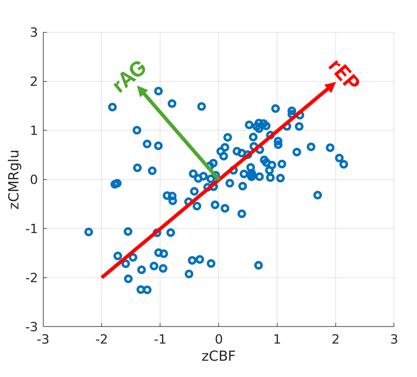
Derivation of rEP and rAG for an example subject.
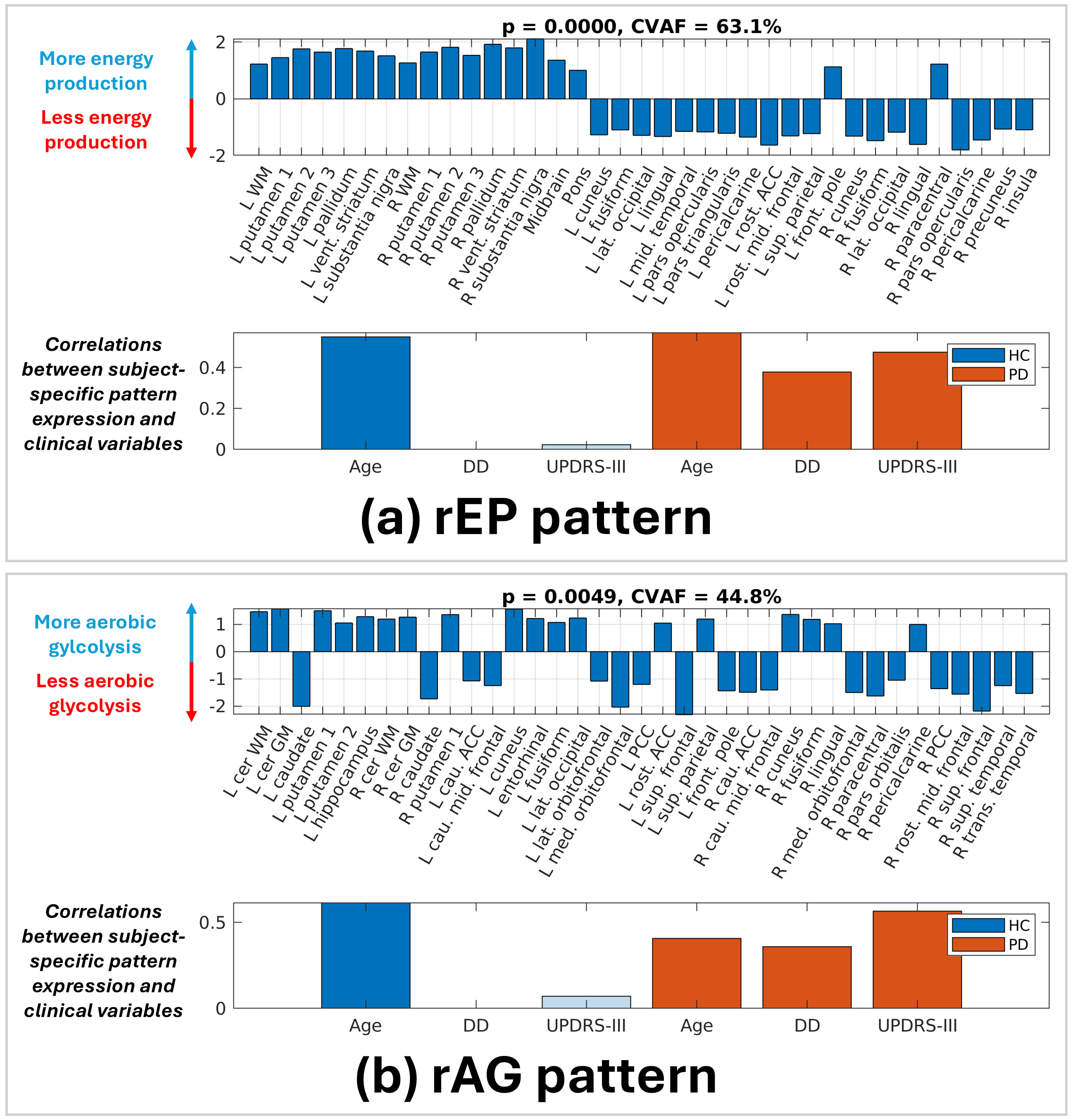
PLSC patterns for rEP and rAG
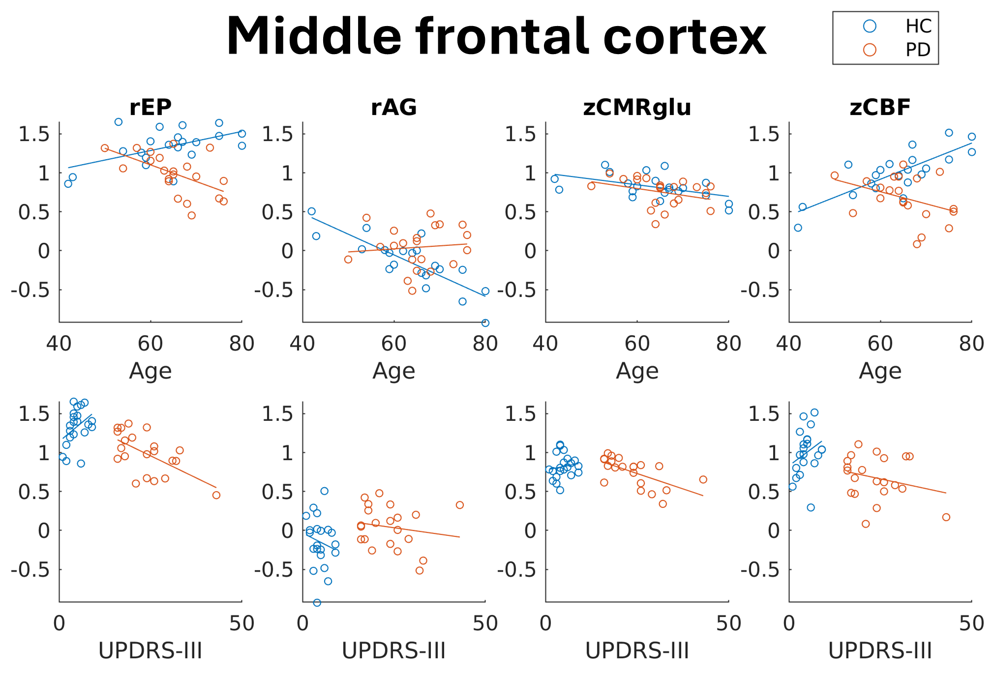
Energetics in midfrontal cortex with trendlines
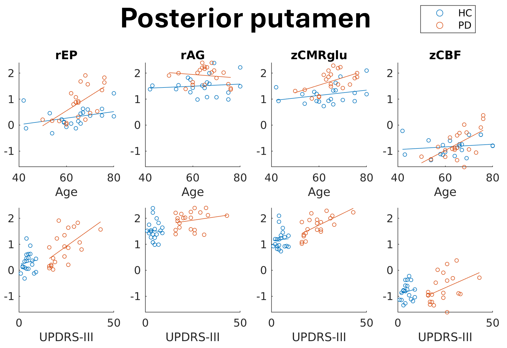
Energetics in posterior putamen with trendlines
References: [1] Ma Y, Tang C, Spetsieris PG, Dhawan V, Eidelberg D. Abnormal metabolic network activity in Parkinson’s disease: test—retest reproducibility. Journal of Cerebral Blood Flow & Metabolism. 2007 Mar;27(3):597-605.
[2] Moeller JR, Ishikawa T, Dhawan V, Spetsieris P, Mandel F, Alexander GE, Grady C, Pietrini P, Eidelberg D. The metabolic topography of normal aging. Journal of Cerebral Blood Flow & Metabolism. 1996 May;16(3):385-98.
[3] Matthews DC, Lerman H, Lukic A, Andrews RD, Mirelman A, Wernick MN, Giladi N, Strother SC, Evans KC, Cedarbaum JM, Even-Sapir E. FDG PET Parkinson’s disease-related pattern as a biomarker for clinical trials in early stage disease. NeuroImage: Clinical. 2018 Jan 1;20:572-9.
[4] Cui H, Kong Y, Zhang H. Oxidative stress, mitochondrial dysfunction, and aging. Journal of signal transduction. 2012;2012(1):646354.
[5] Krishnan A, Williams LJ, McIntosh AR, Abdi H. Partial Least Squares (PLSC) methods for neuroimaging: a tutorial and review. Neuroimage. 2011 May 15;56(2):455-75.
To cite this abstract in AMA style:
C. Bevington, S. Dhaliwal, J. Mckenzie, A. Stoessl, V. Sossi. Brain Energetics: Changes in Healthy Aging and Parkinson’s Disease [abstract]. Mov Disord. 2025; 40 (suppl 1). https://www.mdsabstracts.org/abstract/brain-energetics-changes-in-healthy-aging-and-parkinsons-disease/. Accessed April 7, 2026.« Back to 2025 International Congress
MDS Abstracts - https://www.mdsabstracts.org/abstract/brain-energetics-changes-in-healthy-aging-and-parkinsons-disease/