Session Information
Date: Monday, October 8, 2018
Session Title: Parkinson's Disease: Neuroimaging And Neurophysiology
Session Time: 1:15pm-2:45pm
Location: Hall 3FG
Objective: We aimed to explore whether network connectivity structure as measured by diffusion tensor imaging (DTI) are associated with progression rate of motor and non-motor outcomes in Parkinson’s disease (PD). Secondly, we investigated if brain connectivity measures can differentiate clinical subtypes of PD.
Background: PD is a complex neurodegenerative disorder that varies considerably in its clinical manifestations and prognosis. Investigating biomarkers to predict PD progression, therefore, is of high priority for research and clinical practice.
Methods: This study includes 144 de novo PD patients recruited in the Parkinson’s Progression Markers Initiative (PPMI). A comprehensive set of clinical features including both motor and non-motor symptoms was evaluated at baseline and annual follow-up visits. We used diffusion-weighted MRI (DW-MRI) scans obtained at entry, when participants were at the early drug-naïve stage. Mean diffusivity maps were computed for each participant in six regions of interest in the basal ganglia. For analysis of disease progression, we created a global composite outcome (GCO) as a single numeric indicator of prognosis. We also classified the PPMI population based on multi-domain clinical criteria into three subtypes: ‘mild motor-predominant’, ‘intermediate’ and ‘diffuse malignant’.
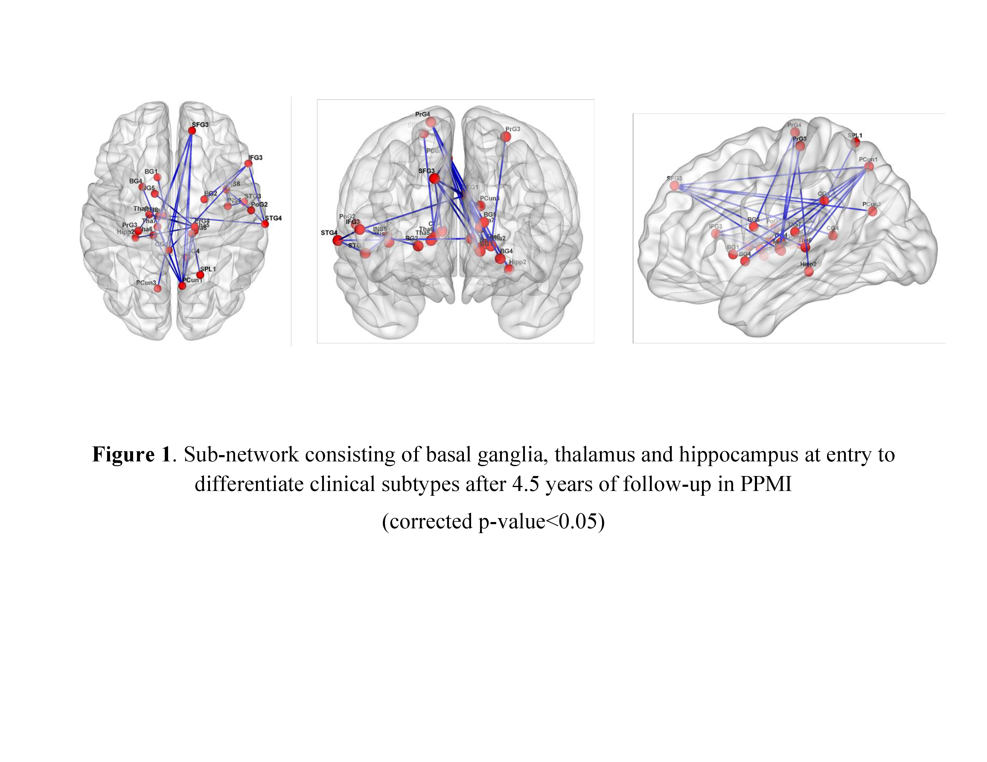
Results: Baseline mean diffusivity of globus pallidus was significantly associated with worsening of motor severity (r=0.27, p=0.001), cognition (r=-0.18, p=0.040), and GCO (r=0.30, p<0.0001) after 4.5 years follow-up. After regressing out the effect of aging, network global efficacy at baseline significantly predicted decline in MoCA (r=0.21, p<0.05), and increase in GCO (r=-0.17, p<0.05). Disruption in a sub-cortical network consisting of basal ganglia, thalamus and hippocampus at baseline was significantly associated with being subtyped as ‘diffuse malignant’ versus ‘mild motor-predominant’ after 4.5 years (corrected p<0.05) [Figure 1].
Conclusions: DW-MRI measures of diffusivity and network connectivity (both whole brain and basal ganglia) demonstrated significant associations with progression of motor and non-motor features, and clinical subtypes of PD after 4.5 years. DW- MRI measures could be used as prognostic biomarkers in clinical trials starting from the early de novo stage of PD.
References: 1. Fereshtehnejad SM, et al (2017). Clinical criteria for subtyping Parkinson’s disease: biomarkers and longitudinal progression. Brain, vol. 140, no. 7, pp. 1959-1976. 2. Zalesky A, et al. (2010). Network-based statistic: identifying differences in brain networks. Neuroimage, vol. 53, no. 4, pp. 1197-207.
To cite this abstract in AMA style:
S.M. Fereshtehnejad, N. Abbasi, Y. Zeighami, K. Larcher, R. Postuma, A. Dagher. Brain Network Connectivity Measured by Diffusion Tensor Imaging Predicts Prognosis in Parkinson’s Disease [abstract]. Mov Disord. 2018; 33 (suppl 2). https://www.mdsabstracts.org/abstract/brain-network-connectivity-measured-by-diffusion-tensor-imaging-predicts-prognosis-in-parkinsons-disease/. Accessed July 18, 2025.« Back to 2018 International Congress
MDS Abstracts - https://www.mdsabstracts.org/abstract/brain-network-connectivity-measured-by-diffusion-tensor-imaging-predicts-prognosis-in-parkinsons-disease/

