Session Information
Date: Monday, June 20, 2016
Session Title: Surgical therapy: Parkinson's disease
Session Time: 12:30pm-2:00pm
Location: Exhibit Hall located in Hall B, Level 2
Objective: This work helps validate the utility of direct targeting using 3T T1 inverted sequences with atlas application during planning for DBS electrode placement. We argue that direct targeting should be the standard of care to ensure precise and accurate DBS electrode placement.
Background: The methods by which DBS electrodes are implanted vary widely amongst surgeons. At the University of Florida we utilize a T1 inverted sequence on a 3T MR to optimally define and target STN, GPi, or the thalamus. A deformable atlas is applied to demarcate the aforementioned structures. To confirm that the structures on our transformed atlas are indeed those we perceive them to be, we imaged post-mortem brains implanted with DBS leads using 17T MR Microscopy. The images provide clear delineation of the basal ganglia substructures and define the location of the implanted DBS leads at 78 um resolution.
Methods: Post-mortem brain specimens from patients who had been implanted with DBS for PD were obtained. The specimens underwent formalin fixation within 24 hours after death. The DBS electrodes were removed after fixation. The brain specimen underwent 3T MR imaging. The atlas was applied per our standard planning technique. The brain was sectioned and the area of interest around the DBS electrode was imaged with a T1 sequence at 78 um resolution using 17T MR Microscopy. The 3T and 17T images were fused.
Results: A 68-year-old man with bilateral STN DBS for PD underwent post-mortem imaging. Fusion of the 3T sequence with the anatomic atlas overlay to the 17T sequence confirmed that basal ganglia structures demarcated on the atlas corresponded with those visualized on the 17T MR microscopy. Figure 1: 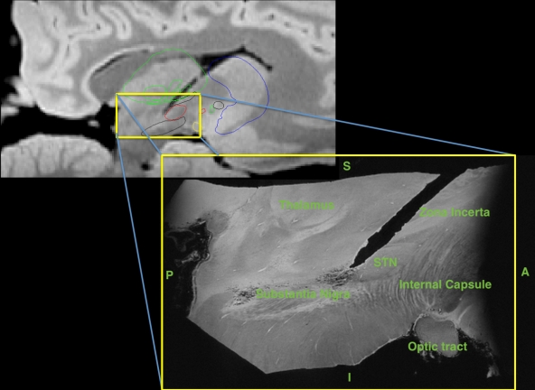 3T and 17T representative sagittal image demonstrating the thalamus, subthalamic nucleus, substantia nigra, zona incerta, internal capsule, optic tract, and DBS lead tract. Figure 2:
 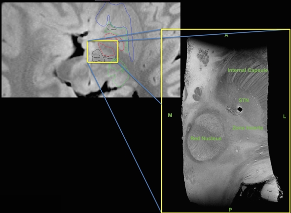 3T and 17T representative axial image demonstrating the red nucleus, subthalamic nucleus, zona incerta, internal capsule, and DBS lead tract.
Conclusions: We have developed a novel method of direct targeting at UF utilizing an inverted T1 3T MRI. We obtained post-mortem 17T MR demonstrating DBS lead placement posterior to the STN, yet having excellent clinical efficacy. This preliminary work supports our use of 3T imaging and an atlas matching technique to define the STN, GPi and thalamus and achieve precise and accurate placement of the DBS lead.
To cite this abstract in AMA style:
J.D. Hilliard, T. Morishita, C.H. Lee, S.J. Blackband, M.S. Okun, K.D. Foote. DBS targeting: Validation of 3T MR image guided targeting using post-mortem 17T MR microscopy [abstract]. Mov Disord. 2016; 31 (suppl 2). https://www.mdsabstracts.org/abstract/dbs-targeting-validation-of-3t-mr-image-guided-targeting-using-post-mortem-17t-mr-microscopy/. Accessed June 18, 2026.« Back to 2016 International Congress
MDS Abstracts - https://www.mdsabstracts.org/abstract/dbs-targeting-validation-of-3t-mr-image-guided-targeting-using-post-mortem-17t-mr-microscopy/