Session Information
Date: Wednesday, June 22, 2016
Session Title: Parkinson's disease: Neuroimaging and neurophysiology
Session Time: 12:00pm-1:30pm
Location: Exhibit Hall located in Hall B, Level 2
Objective: 1. Can brainstem changes be detected using quantitative MRI (qMRI). 2. How many subjects would be required to detecting changes in brainstem regions affected in the earliest stages of Parkinson’s disease (PD).
Background: Brainstem pathology occurs early in PD, yet neuroimaging studies have failed to detect this. This may be due to the difficulties in imaging this region. Previously, we developed a method(1) using qMRI to segment the brainstem into four tissue classes, which is applied here.
Methods: Data Collection: 7 patients with PD (mean age 53.14y) and 7 healthy controls (mean age 55.14y) underwent 3T MRI. Quantitative magnetic transfer (MT), proton density (A), R2s and R1 maps were acquired. T1w images from 223 subjects in the PPMI (152 PD) were downloaded for comparison. Structural Processing
Brainstem pre-processing is described in (1). PPMI data was segmented in SPM12, warped with the Shoot toolbox, and the brainstem region masked. Morphometric Analysis
Two sample T-test were performed in SPM12. Whole brainstem tensor based morphometry (TBM) and voxel based quantification (VBQ) was performed controlling for age, gender and intracranial volume. Sample Size Calculations
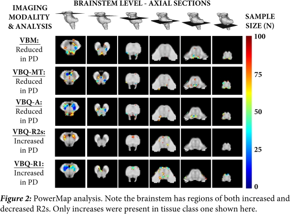
The morphometric analysis was repeated but masked to tissue class one containing all the regions of interest (ROIs). The PowerMap toolbox was used to calculate 90% power using a FWE corrected P < 0.05. The median number of subjects required to detect change within each ROI extracted.
Results: Morphometric changes: Overall asymmetric volumetric reduction in the substantia nigra (SN), associated with raised proton density and reduced MT: 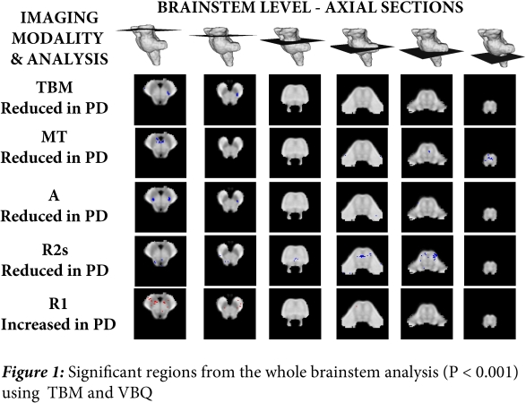 Power Calculations: All brainstem ROIs, selected using histological staging as an a priori, are detectable at modest sample sizes:
| Median Number of Subjects to Detect Regional Structural Changes in Patients at 90% Power and FWE corrected P <0.05 | ||||||||
| IMAGING MODALITY AND DIRECTION OF CHANGE IN PD | ||||||||
| Reduced VBM | Reduced MT | Reduced A | Increased R1 | Increased R2s | Reduced R2s | AVERAGE | ||
| Substantia Nigra | 29 | 63 | 20 | 31 | 70 | – | 43 | |
| Pedenculopontine Nucleus | 32 | 42 | 36 | – | – | 38 | 37 | |
| Raphe Nucleus (Mesencephalon) | 45 | 71 | 56 | – | – | 26 | 50 | |
| Raphe Nucleus (pontine) | 69 | 86 | 56 | – | – | 42 | 63 | |
| Dorsal Motor Nucleus of the Vagus | 56 | 52 | 56 | – | – | 54 | 55 | |
 Comparison with PPMI: No significant regions were detected using TBM. The PowerMap analysis was unable to detect any effect.
Conclusions: Despite the small cohort, this work combined high-resolution qMRI with the brainstem segmentation method, demonstrating brainstem changes in Parkinson’s disease can be detected. In contrast, standard T1w imaging was unable to detect these changes irrespective of sample size. This work has important implications for non-invasive biomarkers of PD and future trial design. REFERENCES: 1. Lambert, C, et al. "Multiparametric brainstem segmentation using a modified multivariate mixture of Gaussians." NeuroImage: clinical 2 (2013): 684-694.
To cite this abstract in AMA style:
C. Lambert, A. Lutti, B. Draganski, T. Foltynie. Detecting brainstem changes in Parkinson’s disease using quantitative MRI [abstract]. Mov Disord. 2016; 31 (suppl 2). https://www.mdsabstracts.org/abstract/detecting-brainstem-changes-in-parkinsons-disease-using-quantitative-mri/. Accessed May 14, 2026.« Back to 2016 International Congress
MDS Abstracts - https://www.mdsabstracts.org/abstract/detecting-brainstem-changes-in-parkinsons-disease-using-quantitative-mri/