Session Information
Date: Wednesday, June 22, 2016
Session Title: Parkinson's disease: Neuroimaging and neurophysiology
Session Time: 12:00pm-1:30pm
Location: Exhibit Hall located in Hall B, Level 2
Objective: To asses white matter tractography of the corticospinal tract in PD patients.
Background: Since the diagnosis of PD is still based on clinical assessment, there is an urgent need to find biomarkers for early diagnosis and prognosis in PD. Previous studies assessed DTI values as a possible biomarker for PD with partial success. Using a bootstrapping algorithm FA values from the whole brain differentiated PD patients from controls with 0.90 AUROC. Nigral fibers tractography also seem to differentiate early PD patients from controls.
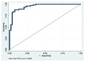
Methods: 58 patients with PD (42 men, mean age 60.3 years, SD 8.99; average disease duration 7.88, SD 6.29) and 40 healthy subjects (mean age 57.8 years, SD 10.59) underwent the same MRI protocol, and 46 patients were assessed by clinical scales and a complete neurological evaluation. We performed DTI analysis using the ExploreDTI software to compare PD patients and controls. For statistical analysis we used Stata 13.1 (http://www.stata.com). Receiver operating characteristics analysis was performed to directly compare groups. Sensitivity and specificity values were quantified for control vs. PD patients.
Results: We found higher FA and reduced medial (MD), radial (RD) and axial diffusivity (AD) in the corticospinal tract. Diffusion values from the mild PD group were also different than controls. FA values higher than 0.559467 differentiates 85.04% of PD patients from controls with 89,04% sensibility of 79.63% specificity (AUROC=0.9536). 
Conclusions: Our data suggest that DTI analysis of the corticospinal tract may be a possible biomarker for PD. It is probable tough that these alterations might be found in other neurodegenerative diseases, including other forms of parkinsonism. However it is still possible to use these values to measure disease progression in PD.
To cite this abstract in AMA style:
R. Guimaraes, B. Campos, L. Campos, L. Piovesana, P. Azevedo, A. D'Abreu, F. Cendes. Diffusion tensor imaging of the corticospinal tract differentiates Parkinson’s disease (PD) patients from controls [abstract]. Mov Disord. 2016; 31 (suppl 2). https://www.mdsabstracts.org/abstract/diffusion-tensor-imaging-of-the-corticospinal-tract-differentiates-parkinsons-disease-pd-patients-from-controls/. Accessed July 18, 2025.« Back to 2016 International Congress
MDS Abstracts - https://www.mdsabstracts.org/abstract/diffusion-tensor-imaging-of-the-corticospinal-tract-differentiates-parkinsons-disease-pd-patients-from-controls/
