Session Information
Date: Wednesday, September 25, 2019
Session Title: Neuroimaging
Session Time: 1:15pm-2:45pm
Location: Les Muses Terrace, Level 3
Objective: To characterize the topography of SNc involvement in PD patients using neuromelanin (NM)-sensitive MRI (NM-MRI).
Background: PD is characterized by the progressive loss of dopaminergic neurons in the SNc [1]. The paramagnetic neuromelanin-iron complex in these neurons appears bright on NM-MRI [1]. Thus, NM-MRI enables assessing SNc changes in the disease. Neurodegeneration occurs nonuniformly across the SNc predominating in the posterior and lateral, but the 3D spatial pattern of SNc loss over time is not well characterized. Here, based on MRI data from 21 HCs and 29 PD patients, we investigated the spatiotemporal pattern of SNc loss in a study-specific and standardized imaging template.
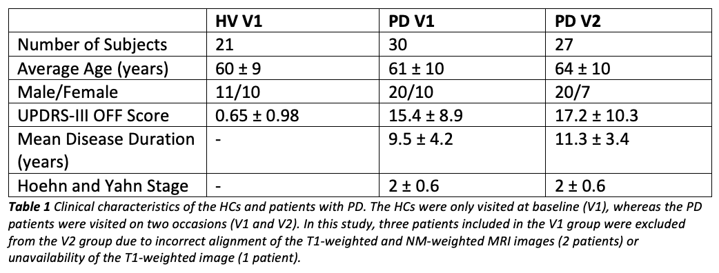
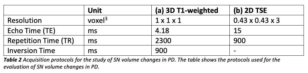
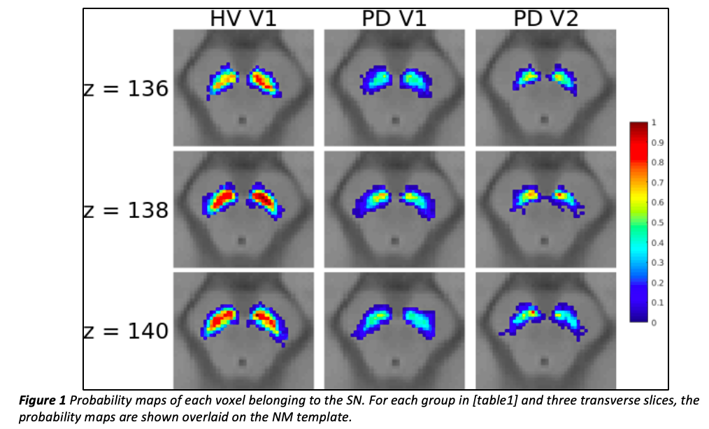
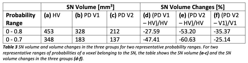
Method: All subjects [table1] were scanned on a Siemens Trio MRI system using a 32-channel head coil, a whole-brain 3D T1-weighted protocol for anatomical reference and an NM-sensitive 2D turbo spin echo protocol with coverage restricted to the midbrain [table2]. Based on the NM-MRI images and for all subjects, a region of interest (ROI) was drawn on the SN by two independent examiners as the hyperintense area dorsal to the cerebral peduncles and ventral to the red nuclei. Based on the T1-weighted images of all subjects, a template of the average brain was calculated using NiftyReg [2,3]. The NM images and SN ROIs of all subjects were aligned to this template. For each group, the coregistered SN ROIs were averaged across subjects to calculate a probability map of each voxel belonging to the SN. These probability maps were thresholded, binarized and used to assess SN volume changes between groups.
Results: In patients, the probability of a voxel belonging to the SN took values across a smaller range than in HVs [figure1] with clusters of low probability localized in the dorsolateral SN. In patients, the SN always had a smaller volume than in the HVs [table3]. These results show quantitatively the spatial pattern of neuronal loss, which has been qualitatively observed to progress from the dorsolateral SNc to more ventral and medial areas of the SN with the progression of disease [1].
Conclusion: Study-specific NM-MRI templates enable noninvasive assessment of spatiotemporal patterns of SN neuronal loss in PD patients vs. HCs. In drug trials, this information could help develop biomarkers of PD progression and modification.
References: 1. Sulzer et al., 2018. Neuromelanin detection by magnetic resonance imaging (MRI) and its promise as a biomarker for Parkinson’s disease. npj Park. Dis. 4, 11. 2. Ourselin et al., 2001. Reconstructing a 3D structure from serial histological sections. Image Vis. Comput. 19, 25–31. 3. Modat et al., 2010. Fast free-form deformation using graphics processing units. Comput. Methods Programs Biomed. 98, 278–284.
To cite this abstract in AMA style:
E. Biondetti, R. Gaurav, L. Yahia-Cherif, G. Mangone, N. Pyatigorskaya, R. Valabregue, C. Ewenczyk, M. Hutchison, JC. Corvol, MJ. Vidailhet, S. Lehéricy. Evaluating Volume Changes in the Substantia Nigra Pars Compacta (SNc) in Parkinson’s Disease (PD) in a Study-Specific Magnetic Resonance Imaging (MRI) Template [abstract]. Mov Disord. 2019; 34 (suppl 2). https://www.mdsabstracts.org/abstract/evaluating-volume-changes-in-the-substantia-nigra-pars-compacta-snc-in-parkinsons-disease-pd-in-a-study-specific-magnetic-resonance-imaging-mri-template/. Accessed March 22, 2026.« Back to 2019 International Congress
MDS Abstracts - https://www.mdsabstracts.org/abstract/evaluating-volume-changes-in-the-substantia-nigra-pars-compacta-snc-in-parkinsons-disease-pd-in-a-study-specific-magnetic-resonance-imaging-mri-template/