Session Information
Date: Monday, October 8, 2018
Session Title: Parkinson's Disease: Neuroimaging And Neurophysiology
Session Time: 1:15pm-2:45pm
Location: Hall 3FG
Objective: To examine the longitudinal changes in neuromelanin (NM) MRI signal in the substantia nigra (SN) in Parkinson’s disease (PD).
Background: PD is characterized by the progressive loss of dopaminergic neurons in the SN pars compacta (SNc), which leads to striatal dopamine depletion. In the striatum, dopamine dysfunction is assessed using radiotracers.1 In the SNc, NM-sensitive MRI showed a reduction in volume and signal that relates to the loss of dopaminergic neurons.2 The longitudinal changes of NM signal in PD are not known.
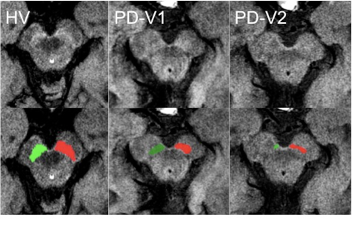
Methods: We prospectively studied 58 idiopathic PD patients at baseline (Visit 1, V1) and follow-up (Visit 2, V2) and 27 age-matched healthy volunteers (HV) at V1 only. Of these subjects, only 29 patients and 23 HV had NM-imaging in all sessions that were free of motion artifacts, included the entire SNc and could be analyzed. The diagnosis of PD was done according to the UK PD brain bank criteria. MRI was performed at 3T (Siemens TRIO) using three-dimensional T1-w images and NM-sensitive images acquired using two-dimensional axial turbo spin echo T1-w images (TR/TE/flip angle: 900ms/15ms/180°, voxel size: 0.4*0.4*3mm3). The regions of interest were traced manually using FreeSurfer in the NM images by two examiners. The SN was delineated as the hyperintense area dorsal to the cerebral peduncle and ventral to the red nucleus (Fig 1). SN signal was normalized by using the background signal intensity of the midbrain at the same level using an in-house software.
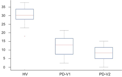
Results: The 29 PD patients (19 men, mean age: 62.1 ± 10.0 years, disease duration: 11.8 ± 4.4 years, Hoehn and Yahn stage: 2.0 ± 0.6, UPDRS-III OFF score: 15.5 ± 9.0 at V1 and 17.3 ± 9.9 at V2) were compared with the 23 HV (12 men, mean age: 59.7±8.3 years, UPDRS-III score: 0.65±0.9). At V1, there was a significant reduction in signal intensity and volume of the SN in PD (12.5 ± 5.6, 160.4 ± 63.7 mm3 respectively) versus HV (30.3 ± 4.6, volume: 251.1 ± 44.5 mm3, p<0.0001, Fig 1 and 2). The patients demonstrated a significant loss in SN signal intensity and volume at V2 (SN signal intensity: 8.6 ± 3.9, SN volume: 115.0 ± 56.2, p<0.0001) as compared to V1 (Fig 1 and 2). The average annual rate of decline was 12.3% for the volume and 12.9 % for signal intensity.
Conclusions: NM-MRI accurately captured longitudinal changes in the SN in PD. Thus, direct noninvasive assessment of SN damage progression in PD seems achievable using NM imaging.
References: 1. de la Fuente-Fernández R, Schulzer M, Kuramoto L, Cragg J, Ramachandiran N, Au WL, Mak E, McKenzie J, McCormick S, Sossi V, Ruth TJ, Lee CS, Calne DB, Stoessl AJ. Age-specific progression of nigrostriatal dysfunction in Parkinson’s disease. Ann Neurol. 2011;69:803-10. 2. Sasaki M, Shibata E, Tohyama K, Takahashi J, Otsuka K, Tsuchiya K, Takahashi S, Ehara S, Terayama Y, Sakai A.Neuromelanin magnetic resonance imaging of locus ceruleus and substantia nigra in Parkinson’s disease. Neuroreport. 2006;17:1215-8.
To cite this abstract in AMA style:
R. Gaurav, N. Pyatigorskaya, C. Ewenczyk, R. Valabregue, K. Evans, I. Arnulf, M. Vidailhet, S. Lehéricy. Longitudinal changes in Neuromelanin MRI Signal in Parkinson’s Disease [abstract]. Mov Disord. 2018; 33 (suppl 2). https://www.mdsabstracts.org/abstract/longitudinal-changes-in-neuromelanin-mri-signal-in-parkinsons-disease/. Accessed March 23, 2026.« Back to 2018 International Congress
MDS Abstracts - https://www.mdsabstracts.org/abstract/longitudinal-changes-in-neuromelanin-mri-signal-in-parkinsons-disease/