Session Information
Date: Monday, October 8, 2018
Session Title: Parkinson's Disease: Neuroimaging And Neurophysiology
Session Time: 1:15pm-2:45pm
Location: Hall 3FG
Objective: To examined quantitative measures of neuromelanin (NM) MRI and DaT-SPECT imaging of the SN and striatum to distinguish PD patients from healthy control subjects and the relationship between imaging modalities.
Background: Parkinson’s disease (PD) is characterized by dopaminergic cell loss within the substantia nigra (SN) and associated loss of nerve terminals in the striatum [1].
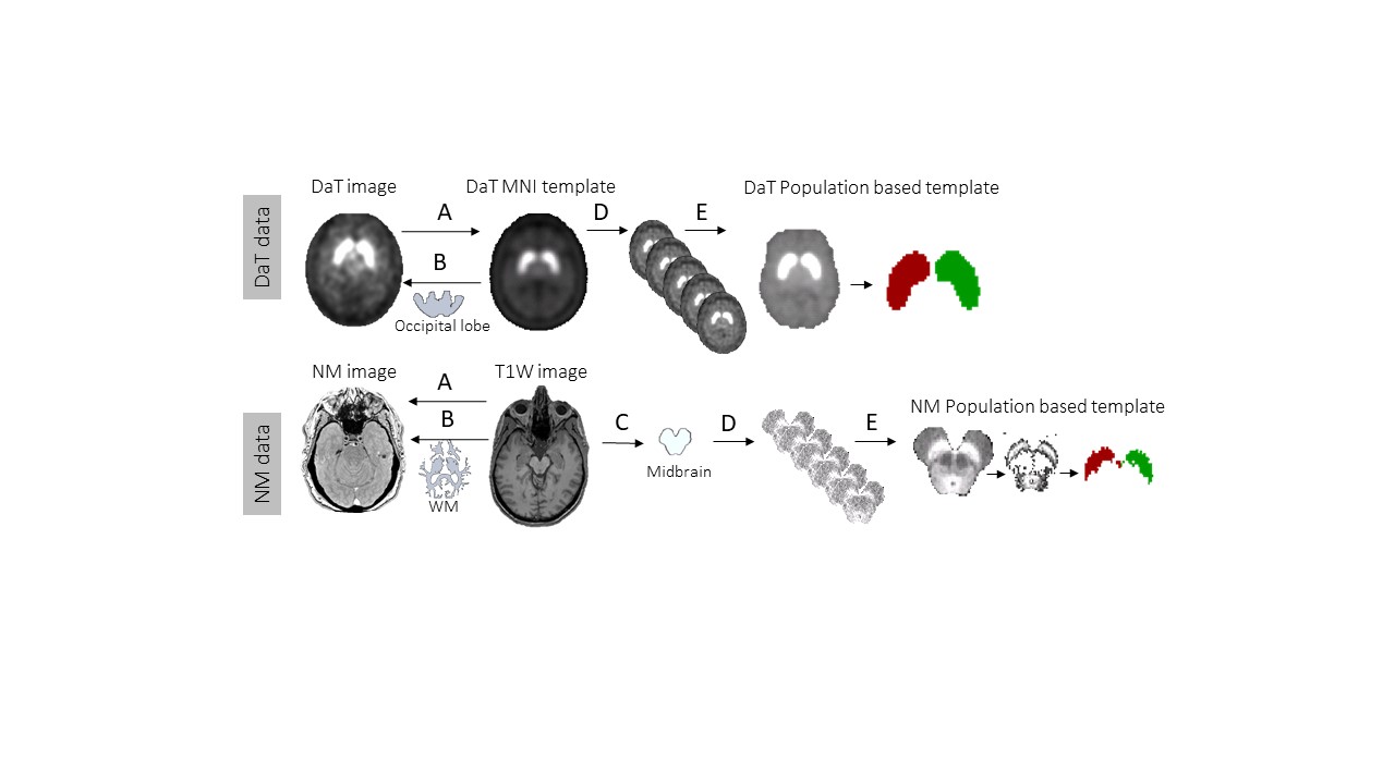
Methods: Subjects: Eight healthy subjects (537yrs) and 20 PD patients (6112yrs) were included. Imaging Data: Striatal SPECT images were acquired via GE Infinia camera 3 hours post IV DaTScanTM 5mCi (185MBq). A 3D-MPRAGE T1 sequence and a gradient echo sequence sensitive to neuromelanin (optimized for SN coverage) were acquired via 3T Siemens Prisma. Image Analysis: Images within each modality were co-registered to a common space (Fig1.A). Intensity normalization was performed relative to occipital lobe (DaT data) and relative to white matter (NM data) (Fig1.B). A population-based template was created for both methods based on the control group. The right and left striatum and SN areas were extracted from each template using intensity and cluster size segmentation (Fig1.C-E). The volume and mean (relative) intensity value of DaT uptake and NM MR signal within the four regions for each subject were measured [figure1].
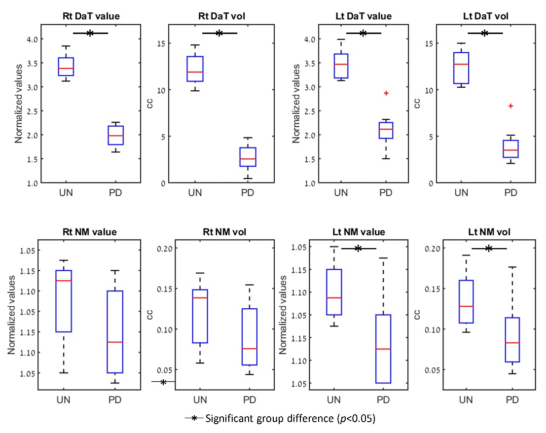
Results: Differences between groups: No significant age or gender differences were detected between groups (p=0.07). Significant group differences (p<0.05) were found for the right and left striatum (DaT) and for the left SN (NM), with greater intensity and volume values detected for the control relative to the PD group [figure2]. Correlations between modalities: Significant positive correlations were detected between the mean relative intensity values (r=0.56, p=0.02) and volumes (r=0.56, p=0.02) of the DaT uptake detected in the left striatum and the NM signal detected in the left SN.
Conclusions: Quantitative analysis of DaT uptake and NM imaging revealed differences between healthy subjects and PD patients. The striatal DaT and SN NM MRI measures were significantly correlated. The findings are consistent with previous work [2] and taken together, support the continued evaluation of NM MRI as an imaging biomarker for PD.
References: 1. Ehringer H & Hornykiewicz O. Parkinsonism Relat. Disord, 1998. 2. Isaias IU, et al. Front Aging Neurosci, 2016.
To cite this abstract in AMA style:
D. Ben Bashat, M. Artzi, A. Mirelman, T. Thaler, A. Orr Urterger, M. Weisz, E. Even-Sapir, K. Evans, R. Hutchison, J. Cedarbaum, H. Lerman Shacham. Quantitative analysis of DaT uptake and neuromelanin MR contrast in patients with Parkinson’s disease and healthy controls [abstract]. Mov Disord. 2018; 33 (suppl 2). https://www.mdsabstracts.org/abstract/quantitative-analysis-of-dat-uptake-and-neuromelanin-mr-contrast-in-patients-with-parkinsons-disease-and-healthy-controls/. Accessed March 24, 2026.« Back to 2018 International Congress
MDS Abstracts - https://www.mdsabstracts.org/abstract/quantitative-analysis-of-dat-uptake-and-neuromelanin-mr-contrast-in-patients-with-parkinsons-disease-and-healthy-controls/