Category: MSA, PSP, CBS: Neuroimaging
Objective: This study evaluates the utility of simple linear measurements of the putamen to distinguish Multiple System Atrophy (MSA) from idiopathic Parkinson’s Disease (iPD) and Progressive Supranuclear Palsy (PSP).
Background: Differentiating MSA from other parkinsonian disorders remains a clinical challenge, particularly at initial presentation.
Method: Two readers retrospectively assessed patients presenting with parkinsonism who underwent 3.0T MRI from 2011-2024 at our institution. Putaminal thickness was measured at the anterior commissure (AC) line and the midline between the AC and posterior commissure (mid AC-PC). Relative measurements were calculated as a percentage of putaminal thickness relative to inner table diameter. Automatic volume estimates of putaminal and total intracranial volume were obtained using FreeSurfer.
Statistical analyses included estimation of intraclass correlation (ICC) coefficients, ROC curve analysis and Pearson correlation coefficients.
Results: The study included 111 patients (35 MSA-P, 11 MSA-mix, 25 PSP, and 40 iPD). No significant differences in age or sex between groups were found. Interobserver reliability was high (ICC >0.8) for all described measurements.
ROC curve analysis demonstrated that decreased putaminal thickness estimated by simple linear measurements is a strong distinguishing feature for MSA patients presenting with parkinsonism compared to PSP and iPD patients. Putaminal diameter at the AC line (AUC 0.92 95%CI [0.87, 0.97]) and mid AC-PC line (AUC 0.94 [0.90, 0.99]) were comparable in discriminatory performance to relative measurements at these levels (AUC 0.93 [0.88, 0.98] and AUC 0.90 [0.84, 0.97] respectively), as well as to automatic volumetric putaminal segmentation (AUC 0.90 [0.83, 0.96]). Best differentiating threshold for MSA diagnosis was less than 8.2 mm at the AC line (87% sensitivity, 80% specificity [figure1]), and less than 3.9 mm at the mid AC-PC line (78% sensitivity, 100% specificity [figure 2]).
Pearson analysis showed moderate to strong positive correlations between volumetric and linear measurements.
Conclusion: The proposed linear metrics with optimal differentiating thresholds assess putaminal atrophy with comparable performance to volumetric metrics. This provides a simple, reliable method to distinguish MSA from other parkinsonian disorders.
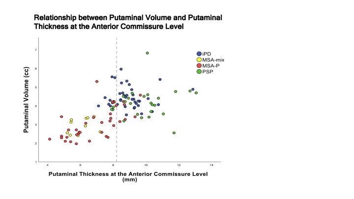
AC level putaminal thickness and putaminal volume
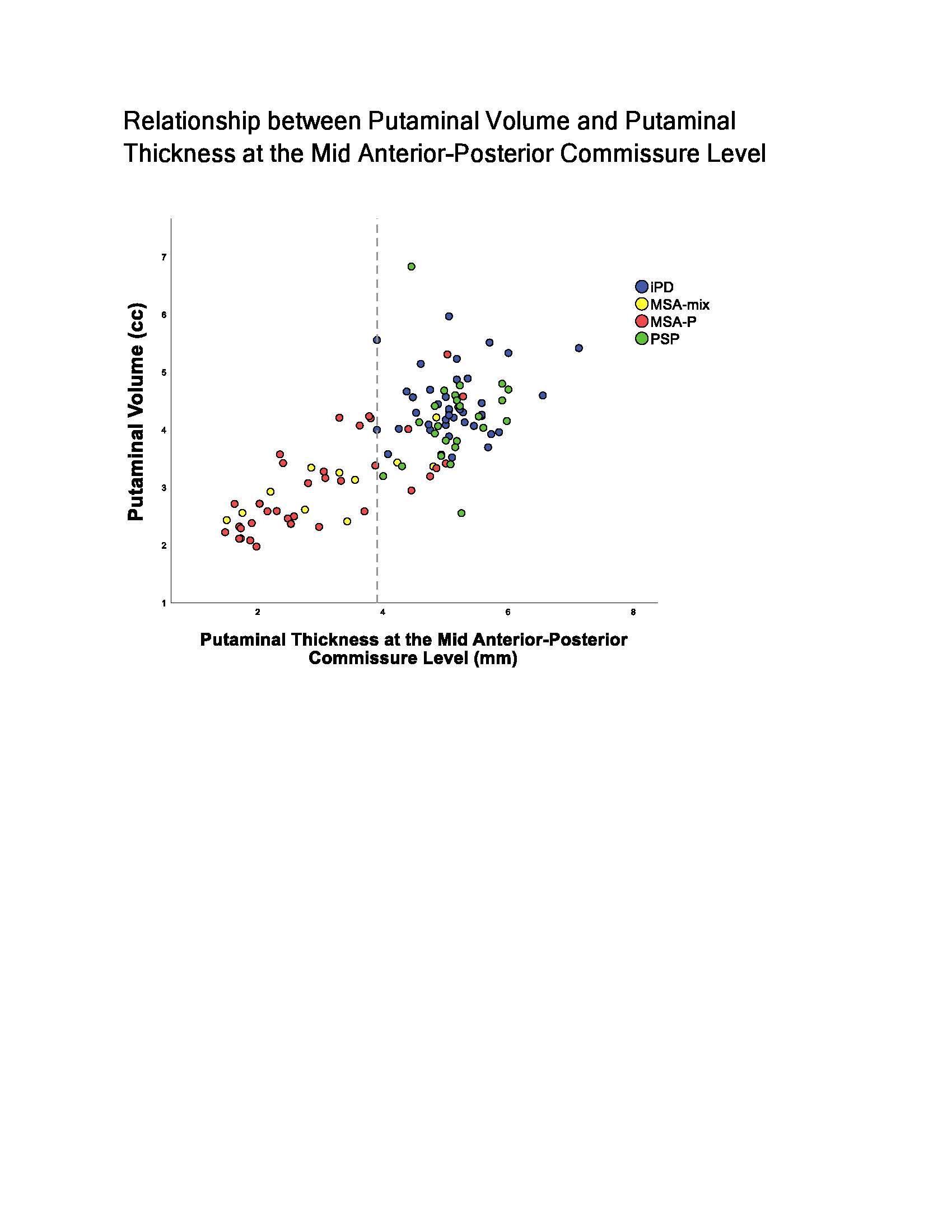
Mid AC-PC putaminal thickness and putaminal volume
To cite this abstract in AMA style:
T. Rohringer, V. Lam Shin-Cheung, P. Alcaide-Leon. Simple linear measurements of the putamen reliably differentiate MSA from other parkinsonian disorders on MRI. [abstract]. Mov Disord. 2025; 40 (suppl 1). https://www.mdsabstracts.org/abstract/simple-linear-measurements-of-the-putamen-reliably-differentiate-msa-from-other-parkinsonian-disorders-on-mri/. Accessed April 7, 2026.« Back to 2025 International Congress
MDS Abstracts - https://www.mdsabstracts.org/abstract/simple-linear-measurements-of-the-putamen-reliably-differentiate-msa-from-other-parkinsonian-disorders-on-mri/