Category: Parkinson's Disease: Neuroimaging
Objective: To determine whether synaptic density differs in the brain of people with Parkinson’s disease and healthy controls using the [18F]-SynVesT-1 PET tracer.
Background: The quantification of synaptic density is a promising diagnostic tool for Parkinsonian disorders. Novel PET radiotracers have been developed to measure synaptic density by targeting the ubiquitously expressed pre-synaptic vesicle protein SV2A. Here we present the preliminary findings of the quantification of synaptic density using [18F]-SynVesT-1 in individuals with Parkinson’s and healthy controls.
Method: 6 subjects with PD (5M/1F) and 9 healthy controls (HC) (2M/7F) underwent a [18F]-SynVesT-1 PET scan on a GE Discovery MI PET-CT. Using Pmod (v4.203), the PET scans were attenuated using the CT scan and aligned to the subjects’ T1-weighted MRI. The non-displaceable binding potential (BPnd) was calculated using a simplified reference tissue model (SRTM2) for the 0-90-minute time window, The centrum semiovale (CS) was used as the reference region due to low specific binding of SV2A (Smart et al., 2023). Group differences in whole brain BPnd and within regions of interest (ROIs) associated with PD were investigated via ANOVA in R studio. Age and sex were included as covariates for all analyses.
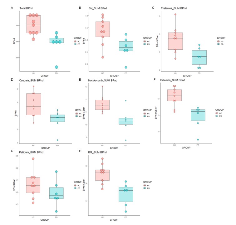
Results: Total brain BPnd was significantly lower in the PD cohort compared to controls (-13.3%) (Figure 1.A). ROI analysis highlighted significant reductions of BPnd in the substantia nigra (-20.6%), thalamus (-17.6%), caudate (-14.9%), nucleus accumbens (-11.4%), putamen (-12.2%), pallidum (-6.6 %) and hippocampus (-12.1%) within the PD cohort (Figure 1.B-G). An average reduction of 12.5 % in BPnd was observed in the basal ganglia (Figure 1.H).
Conclusion: Our results highlight that the quantification of synaptic density via the [18F]-SynVesT-1 tracer is a promising PD diagnostic tool. The reduction in total brain BPnd and ROIs are consistent with previous preclinical and clinical studies quantifying SV2A density in people with PD (Binda et al., 2023; Martin et al., 2023; Wilson et al., 2020). The differences we have observed were independent of age-related change in synaptic density. Importantly, these findings need to be confirmed in a larger sample and investigate the relationship between synaptic density and disease severity.
Figure 1
References: Binda, K., et al (2023). Reduced synaptic SV2A density in a porcine model of Parkinson’s disease and its modulation by deep brain stimulation of the subthalamic nucleus. Brain Stimulation, 252. https://doi.org/10.1016/j.brs.2023.01.406
Martin, S. L., et al (2023). PET imaging of synaptic density in Parkinsonian disorders. Journal of Neuroscience Research. https://doi.org/10.1002/JNR.25253
Naganawa, M., et al (2021). First-in-Human Evaluation of 18F-SynVesT-1, a Radioligand for PET Imaging of Synaptic Vesicle Glycoprotein 2A. Journal of Nuclear Medicine : Official Publication, Society of Nuclear Medicine, 62(4), 561–567. https://doi.org/10.2967/JNUMED.120.249144
Wilson, H., et al (2020). Mitochondrial Complex 1, Sigma 1, and Synaptic Vesicle 2A in Early Drug-Naive Parkinson’s Disease. Movement Disorders : Official Journal of the Movement Disorder Society, 35(8), 1416–1427. https://doi.org/10.1002/MDS.28064
To cite this abstract in AMA style:
S. Martin, C. Uribe, K. Desmond, N. Vasdev, A. Strafella. Synaptic Density Loss in Parkinson’s Disease Independent of Age: Preliminary Findings using [18F]SynVesT-1 PET Tracer [abstract]. Mov Disord. 2024; 39 (suppl 1). https://www.mdsabstracts.org/abstract/synaptic-density-loss-in-parkinsons-disease-independent-of-age-preliminary-findings-using-18fsynvest-1-pet-tracer/. Accessed July 18, 2025.« Back to 2024 International Congress
MDS Abstracts - https://www.mdsabstracts.org/abstract/synaptic-density-loss-in-parkinsons-disease-independent-of-age-preliminary-findings-using-18fsynvest-1-pet-tracer/

