Category: Parkinsonism, Atypical: MSA
Objective: To compare MRI parameters of atrophy in Multiple system atrophy (MSA) subtypes.
Background: MSA is a rare adult-onset synucleinopathology that can be divided in two subtypes depending on whether the prevalence of its symptoms is more parkinsonian or cerebellar (MSA-P and MSA-C respectively). The brain atrophy differences between the subtypes are still controversial.
Method: Thirty-one MSA patients (15 MSA-C and 16 MSA-P) were recruited from hospitals included in the Catalan registry of patients with MSA and transferred to the Parkinson’s disease and Movement Disorders Unit, Hospital Clínic de Barcelona. MSA patients were included following Gilman’s criteria. MRI data were acquired with a 3T scanner (MAGNETOM Trio, Siemens, Germany). FreeSurfer was used to obtain volumetric and cortical thickness measures.
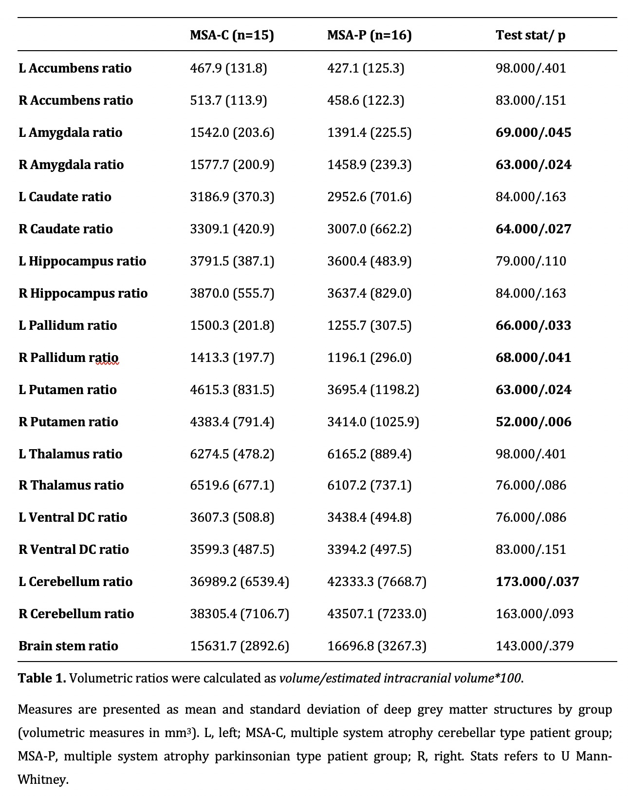
Results: There were no differences in age, sex, years of education, disease duration, motor severity and disease stage. Groups differed in the levodopa equivalent daily dose (p=.001). As for structural data, MSA-C patients had larger atrophy than MSA-P in the left cerebellum, whereas MSA-P showed reduced gray matter volume bilaterally in the pallidum, amygdala and putamen, and right caudate (p<.05 corrected) [table 1]. We did not find differences in measures of global atrophy and cortical thickness.
Conclusion: MSA-C and MSA-P with similar motor impairment and disease duration have structural differences in the cerebellum and deep gray matter structures.
To cite this abstract in AMA style:
A. Campabadal, A. Abos, B. Segura, A. Perez-Soriano, D. Milena, E. Muñoz, Y. Compta, C. Junqué, M. Martí. Deep gray matter atrophy in MSA subtypes [abstract]. Mov Disord. 2021; 36 (suppl 1). https://www.mdsabstracts.org/abstract/deep-gray-matter-atrophy-in-msa-subtypes/. Accessed July 10, 2026.« Back to MDS Virtual Congress 2021
MDS Abstracts - https://www.mdsabstracts.org/abstract/deep-gray-matter-atrophy-in-msa-subtypes/