Category: Neuroimaging (Non-PD)
Objective: This study aims to explore the relationship between left and right putamen volumes in cervical dystonia (CD) patients compared to age-matched healthy controls (HC), while considering age-related variation.
Background: CD is linked to altered putamen volume, yet conflicting data exist regarding its magnitude [1]. Clarifying group-wise volumetric differences is crucial for precise neuroimaging analyses and may shed light on the pathophysiological mechanisms of dystonia.
Method: Structural T1 MPRAGE MRI images were acquired from CD patients (n=23, aged 37-81, mean= 62.7) and age and gender-matched controls. Putamen volumes were derived from segmented volumetric brain images (FreeSurfer 7.3, manually edited), and a general linear model (GLM) analysis assessed age and group (CD vs. HC) effects on corrected left and right putamen volumes.
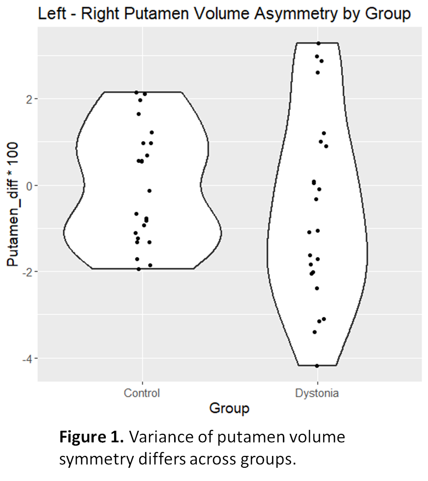
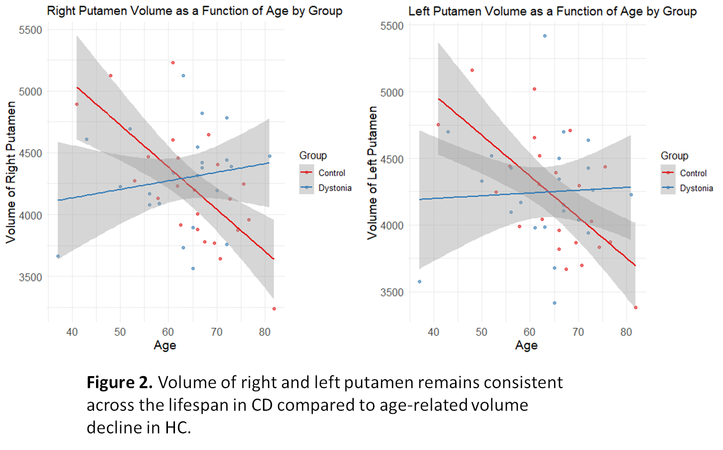
Results: Mean putamen volume did not differ between groups (left, p= 0.97; right, p= 0.72). A Levene’s test confirmed nonhomogeneity of variance across groups (F value= 4.25, p= 0.045), indicating a larger degree of putamen volume asymmetry in participants with CD compared to HC [figure1]. GLM revealed a significant age-group interaction effect on the left (F-statistic= 4.013, p= 0.013) and right (F= 5.82, p= 0.002) putamen volume, with controls showing a steeper decline over the lifespan [figure2]. Framewise displacement did not correlate with the variability of putamen volume in HC (r = 0.28, p = 0.196, df = 21). There was no relationship between framewise displacement and variability of putamen volume in the CD group (Spearmans rho= 0.621, p= 0.11). Right putamen volumes exhibited a more pronounced decline in controls compared to CD (F= 5.816, p= 0.002).
Conclusion: CD is associated with higher variability in putamen volume asymmetry compared to HC, unaffected by interscan motion. The loss of the negative age-volume relationship [2] in CD suggests distinctive pathophysiological changes. This study underscores the importance of further elucidating these mechanisms and exercising caution when implying the putamen as a region of interest in neuroimaging studies.
Figure 1.
Figure 2.
References: 1. MacIver et al, “Structural magnetic resonance imaging in dystonia: A systematic review of methodological approaches and findings” Eur J Neurol, 29(11), 2022, 3418-3448, PMID: 3578541
2. Dima, Danai, et al. “Subcortical Volumes across the Lifespan: Data from 18,605 Healthy Individuals Aged 3–90 Years.” Human Brain Mapping, 43(1), 2022, 452-469, PMID: 33570244
To cite this abstract in AMA style:
M. Owusu-Ansah, A. Tatenbaum, N. Goldman, SA. Norris. Higher Putamen Volume Asymmetry with Age-Preserved Volume in Isolated Adult-Onset Cervical Dystonia [abstract]. Mov Disord. 2024; 39 (suppl 1). https://www.mdsabstracts.org/abstract/higher-putamen-volume-asymmetry-with-age-preserved-volume-in-isolated-adult-onset-cervical-dystonia/. Accessed July 12, 2025.« Back to 2024 International Congress
MDS Abstracts - https://www.mdsabstracts.org/abstract/higher-putamen-volume-asymmetry-with-age-preserved-volume-in-isolated-adult-onset-cervical-dystonia/


