Session Information
Date: Wednesday, June 22, 2016
Session Title: Imaging and Neurophysiology
Session Time: 12:00pm-1:30pm
Objective: To test a novel multimodal MRI approach for in vivo assessment of the subregion of substantia nigra pars compacta (SNpc) most vulnerable to Parkinson’s disease (PD) associated neurodegeneration.
Background: Fearnley and Lees’ 1991 histopathology study of the SNpc in PD famously identified its lateral ventral tier as selectively vulnerable to PD associated death of neuromelanin containing dopamine neurons. Other histopathology studies have identified substantia nigra pars reticulata (SNr) as rich in iron in controls, and SNpc as a site of iron accumulation in PD. MRI pulse sequences have been developed that are sensitive to either iron or neuromelanin. NM-MRI hyperintense signal has been shown to spatially co-localize with areas of neuromelanin containing neurons in radiologic / histologic correlation studies of SN and LC. Our group has identified the region of overlap between iron sensitive susceptibility weighted imaging (SWI) contrast in substantia nigra (SN) in PD subjects and the region of neuromelanin MRI (NM-MRI) contrast in SN in young healthy subjects as spatially consistent with the anatomic location of the lateral ventral tier of SNpc (Langley et al, 2015). We have developed a novel standard space mask to automatically segment this "overlap" region and hypothesize that assessment of neuromelanin contrast in this region will robustly differentiate patients with PD from older controls.
Methods: Two cohorts were used in this abstract. The first consisted of 82 subjects (64 PD, 28 CO) and was acquired with a susceptibility weighted imaging (SWI) sequence. The second consisted of 41 subjects (22 PD, 19 CO) and was acquired using a NM-MRI pulse sequence. All data were acquired with 3T scanners. A standard space neuromelanin SN mask previously reported (Langley et al, 2015) was used to define the NM-MRI SN volume and the overlap between this mask and an iron sensitive SN atlas from the first cohort was used to define our ROI for analysis of NM-MRI signal. Figure 1 shows the overlap region in yellow/red colors. This area appears spatially consistent with the lateral ventral portion of SNpc. 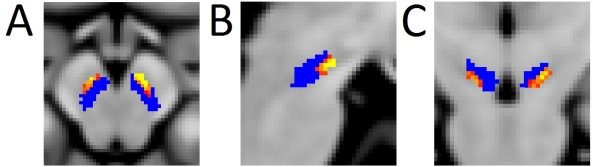
Results: Results are shown in Figure 2 for A) mean MTC in SNpc and B) mean MTC in overlapping region between neuromelanin SN atlas and iron SN atlas. 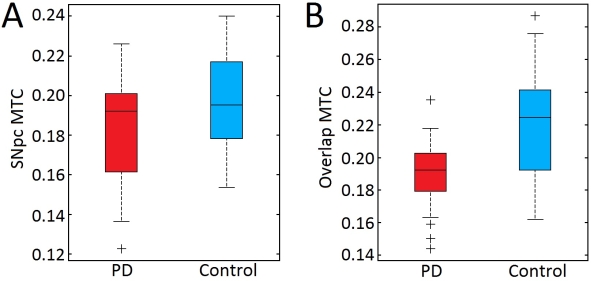
Conclusions: NM-MRI signal in the iron/neuromelanin overlap region in substantia nigra differentiates PD from controls robustly.
To cite this abstract in AMA style:
D.E. Huddleston, J. Langley, J. Sedlacik, K. Boelmans, S.A. Factor, X. Hu. In vivo MRI detection of Parkinson’s disease associated degeneration in the lateral ventral tier of substantia nigra pars compacta [abstract]. Mov Disord. 2016; 31 (suppl 2). https://www.mdsabstracts.org/abstract/in-vivo-mri-detection-of-parkinsons-disease-associated-degeneration-in-the-lateral-ventral-tier-of-substantia-nigra-pars-compacta/. Accessed June 17, 2026.« Back to 2016 International Congress
MDS Abstracts - https://www.mdsabstracts.org/abstract/in-vivo-mri-detection-of-parkinsons-disease-associated-degeneration-in-the-lateral-ventral-tier-of-substantia-nigra-pars-compacta/