Category: Surgical Therapy: Parkinson's Disease
Objective: To compare DBS programming results by novel neuroimaging software assisted programming approach versus standard of care approach in PD patients.
Background: Traditional DBS programming can be time consuming and exhausting to PD patients. It involves withholding anti-parkinsonian medications and a detailed monopolar contact screen.
It is anticipated that a neuroimaging assisted programming software can offer information on electrode placement and simulated electrical field stimulation. In turn, it may facilitate the procedure of DBS programming.
Method: This is a retrospective review conducted at Queen Elizabeth Hospital, Hong Kong. It compares DBS programming result obtained from neuroimaging software assisted programming approach with that from standard clinical approach.
PD patients with neurostimulators implanted between 2018 and 2019 were recruited.
Exclusion criteria included electrode misplacement and abnormal electrode impedance.
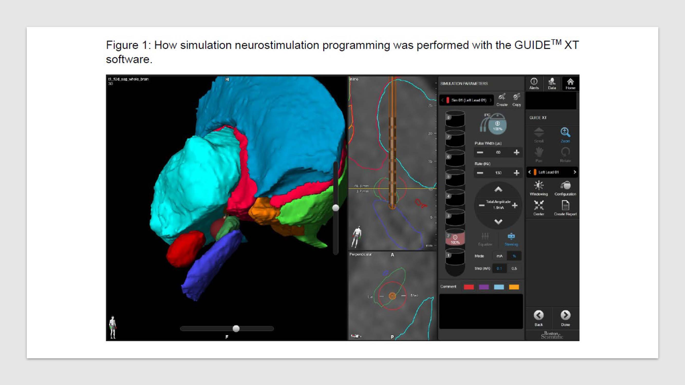
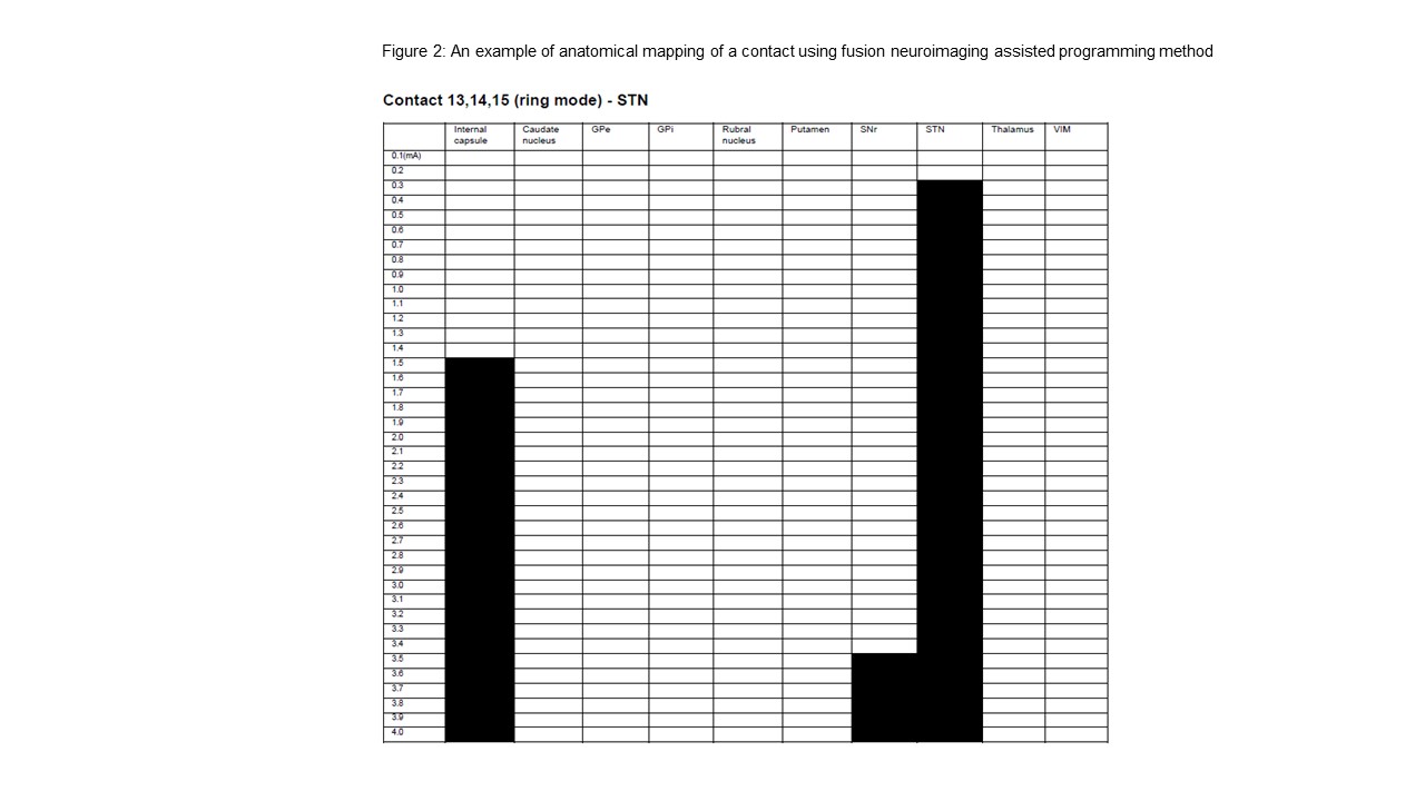
In this study, GUIDETM XT assisted programming (Figure 1) was used. The pre-operative magnetic resonance imaging (MRI) of the brain was first fused with the post-operative computed tomography (CT) of the brain. An automated algorithm located the DBS leads in the post-operative CT scan and provided a 3D reconstruction of the leads and their adjacent structures with the use of Morel atlas registered to the pre-operative MRI. Simulated neurostimulation programming was then performed in every contact. With that, each contact was mapped in details (Figure 2) and so the best contacts were selected.
The results obtained by these two methods were compared using Chi-Square test.
Statistical analyses were performed using SPSS version 20. Statistical significance was set at P < 0.05.
Results: 14 patients were recruited. 1 patient was excluded because of suboptimal electrode location.
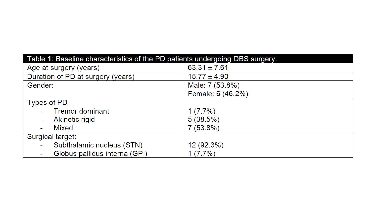
Baseline characteristics are summarised in Table 1.
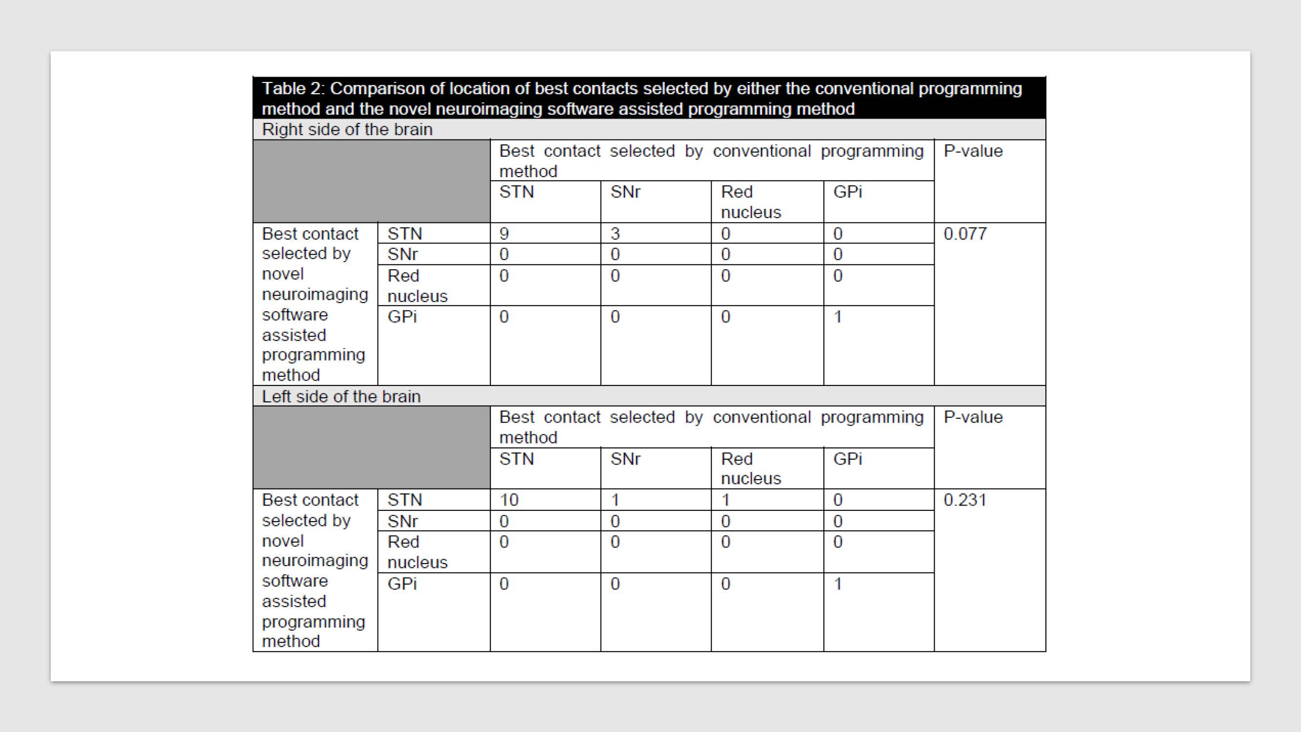
No significant difference was found in the location of best contacts selected by either fusion neuroimaging assisted programming approach or conventional programming approach (Table 2).
Causes of the discrepancy in best contact selection included less robust clinical response (30.8%) and side effects caused by current spread to adjacent structures (7.7%).
Conclusion: This study reported that neuroimaging assisted programming approach was useful in identifying clinically effective contacts and so may be helpful in DBS programming in PD patients.
To cite this abstract in AMA style:
H.F Chan, Y.F Cheung, T.L Poon, M.H Yuen, F.C Cheung, W.L Poon, W.C Fong. Neuroimaging Software Assisted Deep Brain Stimulation (DBS) Programming in Patients with Parkinson’s Disease (PD) [abstract]. Mov Disord. 2020; 35 (suppl 1). https://www.mdsabstracts.org/abstract/neuroimaging-software-assisted-deep-brain-stimulation-dbs-programming-in-patients-with-parkinsons-disease-pd/. Accessed June 30, 2025.« Back to MDS Virtual Congress 2020
MDS Abstracts - https://www.mdsabstracts.org/abstract/neuroimaging-software-assisted-deep-brain-stimulation-dbs-programming-in-patients-with-parkinsons-disease-pd/


