Session Information
Date: Tuesday, September 24, 2019
Session Title: Parkinsonisms and Parkinson-Plus
Session Time: 1:45pm-3:15pm
Location: Agora 3 West, Level 3
Objective: The aim of our study was to determine the diagnostic value of nigral iron depositions in PD, measured by iron specific MRI-techniques.
Background: Nigral iron deposition is considered as an important trigger for oxidative stress and neuro-degeneration. Elevated nigral iron load has been described in Parkinson’s disease (PD) patient’s in histological and imaging analyses.
Method: We included 109 patients with clinically diagnosed PD (79 M/ 30 F; age 63. 7±9.9 years; mean disease duration 4.7±5.2 years) and 109 age-matched healthy controls (HC). All subjects underwent a 3T cerebral MRI protocol, as well as detailed clinical examinations. To measure nigral iron load we performed R2*-relaxometry and Quantitative Susceptibility Mapping (QSM) in total substantia nigra (SN) as well as in pars compacta (SNc) and pars reticulata (SNr) separately. Regions of interest were drawn manually on an iron independent contrast. Group differences were calculated by t-test (for normally distributed variables), otherwise by Mann-Witney-test. For determination of diagnostic value, we performed ROC-analyses and for clinical correlations Spearman correlations.
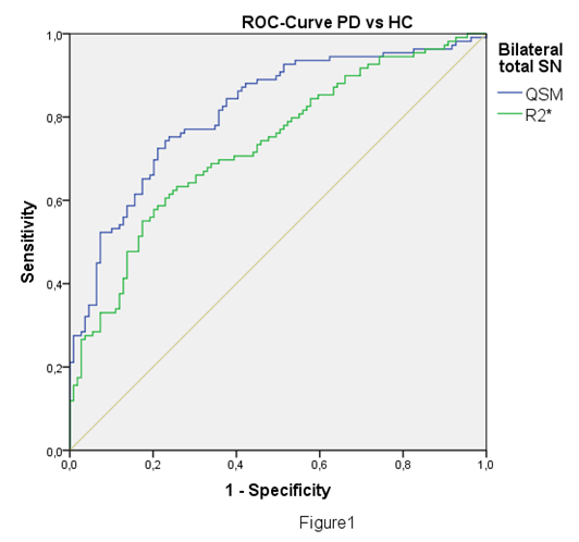
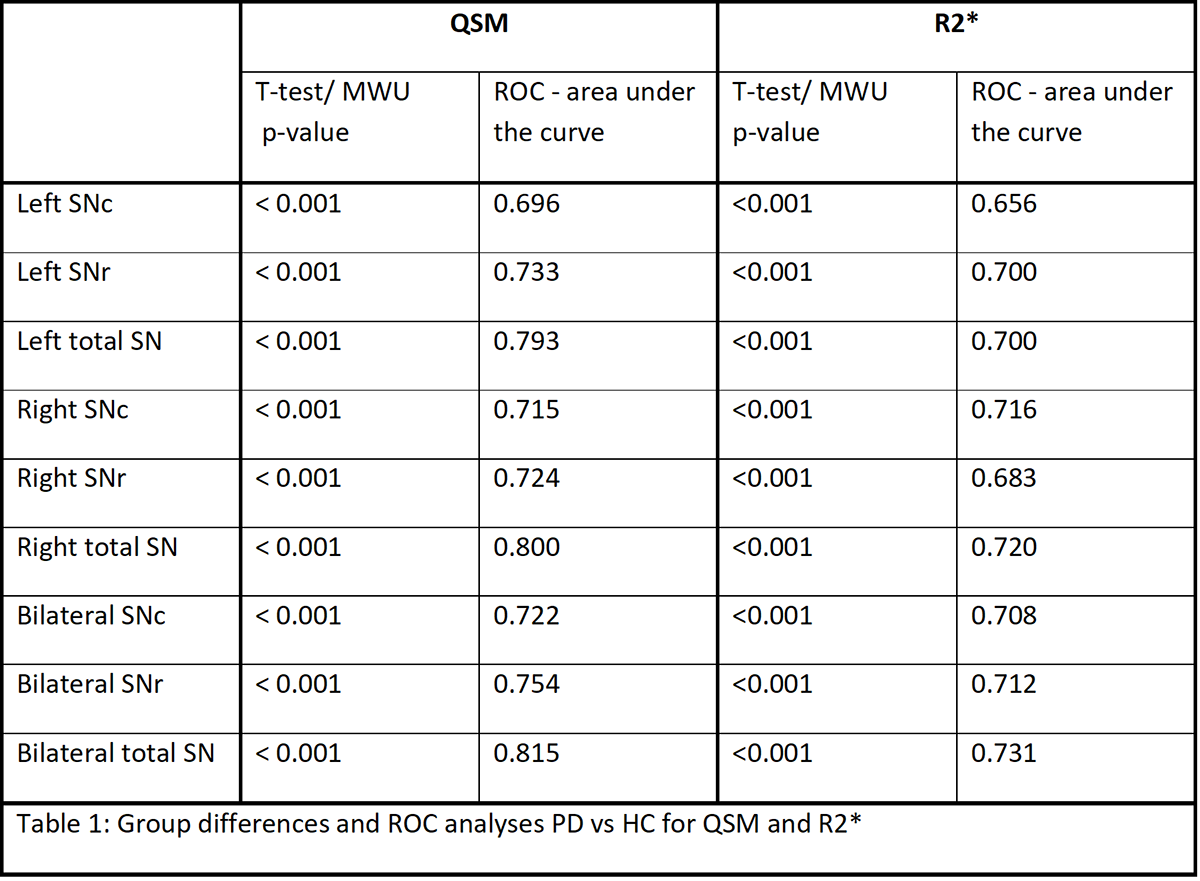
Results: In PD QSM and R2* values were significantly higher with a p-value <0.001 in each region (left, right and bilateral SN, SNc and SNr).Bilateral total SN showed the best group discrimination, for QSM there was an area under the curve of 0.815, a sensitivity of 76.1% and a specificity of 73.4%. For R2* we found an area under the curve of 0.731, a sensitivity of 67.9% and specificity of 67.0% (for detailed results see Table1 and Figure1). For QSM and R2* we found moderate positive correlations between SN and Nonmotor Symptoms Scale and UPDRS.
Conclusion: We found significantly higher iron load in SN in PD compared to HC and positive correlations with parkinsonian motor and nonmotor symptoms. QSM was superior to R2* considering diagnostic sensitivity and showed very good group discrimination. There were no relevant side-differences in nigral iron load and total SN showed better discrimination compared to the sub-regions. QSM values for bilateral total SN showed the highest sensitivity and specificity combination and might be used as diagnostic marker.
To cite this abstract in AMA style:
S. Franthal, L. Pirpamer, N. Homayoon, M. Koegl, P. Katschnig-Winter, K. Wenzel, C. Langkammer, S. Ropele, F. Fazekas, R. Schmidt, P. Schwingenschuh. Nigral Iron Load as Diagnostic Parameter in Parkinson’s Disease [abstract]. Mov Disord. 2019; 34 (suppl 2). https://www.mdsabstracts.org/abstract/nigral-iron-load-as-diagnostic-parameter-in-parkinsons-disease/. Accessed July 5, 2025.« Back to 2019 International Congress
MDS Abstracts - https://www.mdsabstracts.org/abstract/nigral-iron-load-as-diagnostic-parameter-in-parkinsons-disease/


