Category: Parkinson's Disease: Neuroimaging
Objective: Explore the relationship between MDS-UPDRS motor sub-scores (MDS-UPDRS-III) and cerebral perfusion in different Regions of Interest (ROI) in patients with Parkinson’s disease (PwP).
Background: Cerebral blood flow (CBF) using perfusion magnetic resonance imaging (MRI) is under investigation as a potential biomarker for Parkinson’s disease (PD). Studies have shown widespread cortical hypoperfusion and subcortical hypoperfusion in the caudate of PwP[1]. Asymmetry effects related to regional perfusion changes have been described in PD, especially in the putamen, globus pallidus, and thalamus [2]. We hypothesize that the severity of motor symptoms may correlate with perfusion in ROI in PwP.
Method: Baseline data was analyzed from PwP recruited to participate in a phase II trial for PD[3]. Imaging from 16 different subcortical ROI was collected in an OFF-medicine state using a 3 Tesla Philips Ingenia MR scanner. Cerebral perfusion was assessed using a pseudo-continuous arterial spin labeling (pCASL) MRI sequence with gradient-echo echo-planar imaging. Correlations between MDS-UPDRS-III and CBF were assessed in a linear regression analysis with age, sex, and duration of disease (DoD) as independent variables, with a Bonferroni corrected p-value of 0.05. The ROI statistical tests were carried out using SPSS 15.
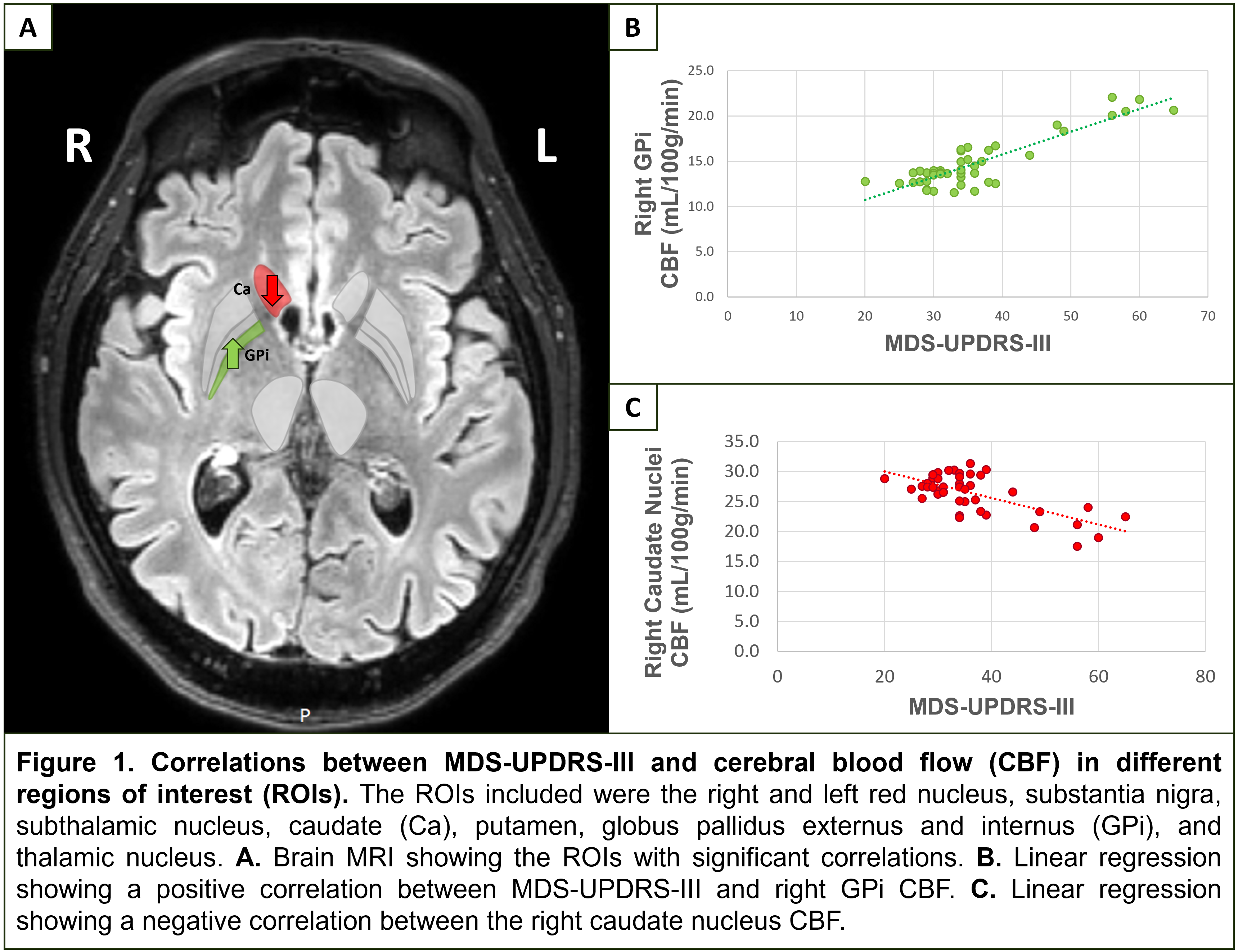
Results: 44 patients were diagnosed with mild to moderate PD; 77.3% were male and 54.5% exhibited right-predominant symptoms. Mean age was 67 years (SD±7, range: 50-80), with a DoD averaging 5 years (SD±2, range: 2-9). Mean MDS-UPDRS-III score was 36 points (SD±10, range: 20-65). Among the 16 ROI, the overall regression was statistically significant in the right Globus Pallidus internus (GPi) (R2 = 21.0, F(4, 38) = [2.533], p = 0.05) and right caudate nuclei (R2 = 21.6, F(4, 38) = [2.619], p = 0.05). It was found that MDS-UPDRS-III had a significantly positive linear association in the right GPi (β = 0.299, p = 0.025) (figure1b), and a negative association in the right caudate nuclei (β = -0.302, p = 0.045) (figure1c).
Conclusion: In this selective PwP population, our findings support that asymmetry of perfusion can occur. We found that patients with more severe motor symptoms have a linear increase in CBF in the right GPi, most likely related to its hyperactivity. In contrast, CBF in the caudate nucleus was decreased, which could be associated with its hypoactivity.
Figure 1
References: 1. Fernandez-Seara MA, Mengual E, Vidorreta M, Aznarez-Sanado M, Loayza FR, Villagra F, Irigoyen J, Pastor MA. Cortical hypoperfusion in Parkinson’s disease assessed using arterial spin labeled perfusion MRI. Neuroimage. 2012;59(3):2743-50. Epub 20111018. doi: 10.1016/j.neuroimage.2011.10.033. PubMed PMID: 22032942.
2. Shang S, Wu J, Zhang H, Chen H, Cao Z, Chen YC, Yin X. Motor asymmetry related cerebral perfusion patterns in Parkinson’s disease: An arterial spin labeling study. Hum Brain Mapp. 2021;42(2):298-309. Epub 20201005. doi: 10.1002/hbm.25223. PubMed PMID: 33017507; PMCID: PMC7775999.
3. ClinicalTrials.gov. Phase IIa Randomized Placebo Controlled Trial: Mesenchymal Stem Cells as a Disease-modifying Therapy for iPD. Available from: https://clinicaltrials.gov/study/NCT04506073.
To cite this abstract in AMA style:
J. Martinez-Lemus, E. Tharp, T. Ellmore, C. Adams, R. Abuamouneh, E. Rodarte, M. Schiess. Severity of Motor Symptoms Correlates with Asymmetric Changes in Globus Pallidus Internus and Caudate Nuclei in Parkinson’s Disease [abstract]. Mov Disord. 2024; 39 (suppl 1). https://www.mdsabstracts.org/abstract/severity-of-motor-symptoms-correlates-with-asymmetric-changes-in-globus-pallidus-internus-and-caudate-nuclei-in-parkinsons-disease/. Accessed July 12, 2025.« Back to 2024 International Congress
MDS Abstracts - https://www.mdsabstracts.org/abstract/severity-of-motor-symptoms-correlates-with-asymmetric-changes-in-globus-pallidus-internus-and-caudate-nuclei-in-parkinsons-disease/

