Category: Ataxia
Objective: To assess substantia nigra pars compacta (SN) degeneration in SCA type 2 and 7 using neuromelanin-sensitive MRI
Background: Spinocerebellar ataxias (SCA) are often associated with extracerebellar signs including parkinsonian symptoms. Neuromelanin-sensitive MRI is a non-invasive tool for studying SN degeneration
Method: The population included SCA mutation carriers (15 SCA2,15 SCA7), and ten age- and sex-matched healthy controls (HC). Subjects were classified as preataxic for SARA (Scale for the Assessment and Rating of Ataxia)<3 and as ataxic for SARA≥3. Other measurements included Inventory of Non-Ataxia Signs (INAS), Cerebellar Cognitive Affective Syndrome Scale (CCAS), an emotional recognition scale, and plasma neurofilament light chains (NfL).
Using a Prisma Siemens 3T system, the MRI protocol included a T1 MPRAGE sequence 1-mm isotropic and a T1 turbo spin-echo sequence for neuromelanin-sensitive imaging 3 (voxel size, 0.4×0.4x3mm3).
The SN was manually segmented to derive the SN volume and signal-to-noise ratio (SNR). The pons volume was extracted after automatic segmentation of T1 images using FreeSurfer6.0
Results: The clinical characteristics are provided in [Table 1].
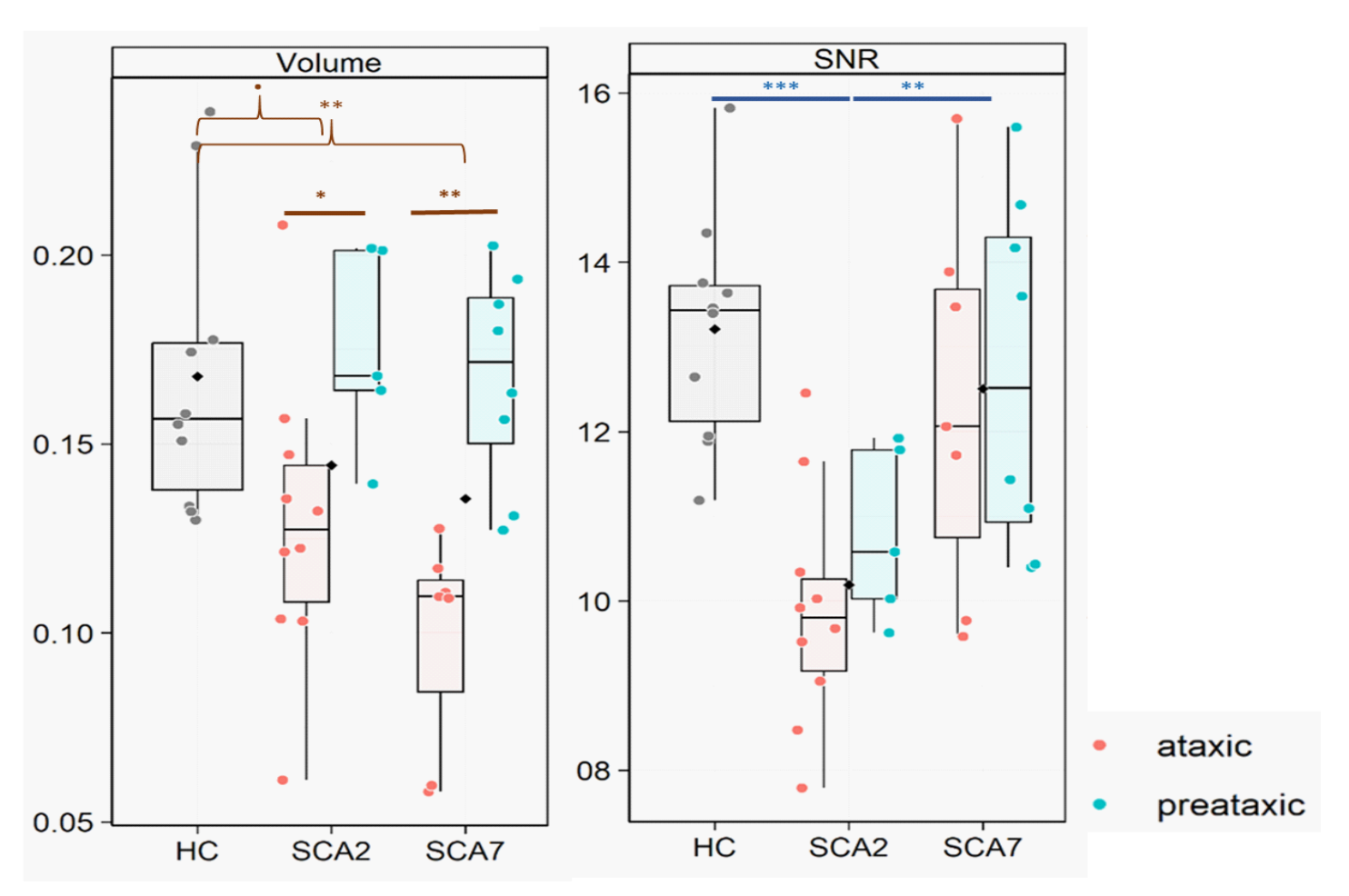
Subjects with SCA2 had lower SNR compared to HC (p=0.0003) and SCA7 subjects (p=0.003 and p=0.03). SNR values did not differ between SCA7 subjects and HC. Preataxic subjects did not differ from HC in both SCA group ([Figure 1]). SCA2 (p=0,0016) and SCA7 (p=0,0022) subjects had lower pons volume than HC.
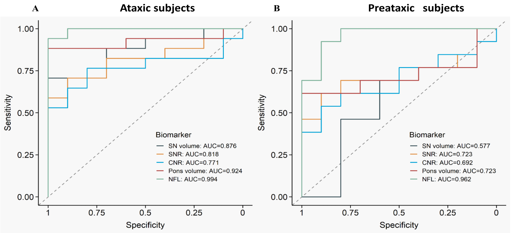
Pons volume had the highest AUC for discriminating HC from ataxic subjects (AUC:0.924), similar to NfL (AUC:0.994). For the differentiation of preataxic subjects, pons volume (AUC:0.723) and SNR (AUC:0.723) both yielded a moderate performance, lower than NfL (AUC:0.962; p=0.07 and p=0.03, respectively) ([Figure 2]).
Considering all SCA subjects, there was a negative correlation between SN volume and time to onset (Pearson coefficient=-0.559; p=0.008), SARA (-0.659; p=0.001) and NfL (-0.464; p=0.027), and a positive correlation with CCAS (0.457; p=0.027) and the emotional recognition scale (0.521; p=0.013)
Conclusion: Neuromelanin-sensitive imaging provides evidence of nigral degeneration in SCA type 2 and 7, that are correlated with disease severity scores and NfL levels. Future studies on larger samples and longitudinal datasets will allow to identify this modality as biomarker even in preataxic stages of the disease
Table1
Figure1
Figure2
To cite this abstract in AMA style:
L. Chougar, G. Coarelli, FX. Lejeune, R. Gaurav, P. Ziegner, A. Durr, S. Lehéricy. Substantia Nigra Degeneration In Spinocerebellar Ataxia 2 And 7 Using Neuromelanin-Sensitive Imaging [abstract]. Mov Disord. 2024; 39 (suppl 1). https://www.mdsabstracts.org/abstract/substantia-nigra-degeneration-in-spinocerebellar-ataxia-2-and-7-using-neuromelanin-sensitive-imaging/. Accessed July 1, 2025.« Back to 2024 International Congress
MDS Abstracts - https://www.mdsabstracts.org/abstract/substantia-nigra-degeneration-in-spinocerebellar-ataxia-2-and-7-using-neuromelanin-sensitive-imaging/


