Session Information
Date: Monday, June 5, 2017
Session Title: Surgical Therapy: Other Movement Disorders
Session Time: 1:45pm-3:15pm
Location: Exhibit Hall C
Objective: We investigated Volumes of Tissue activated (VTA) in dystonia subjects under DBS. We aimed to disentangle the SweetSpot for dystonia suppression within the pallidal region.
Background: GPi-DBS is an established therapy for generalized and cervical dystonia. Average improvement of dystonia severity amounts to 50-60%, but outcomes are often variable and studies report up to 25% non-responders. Variability in electrode placement may account for a large proportion of outcome variability. So far no study has been able to identify an “optimal efficacy volume” within the GPi.
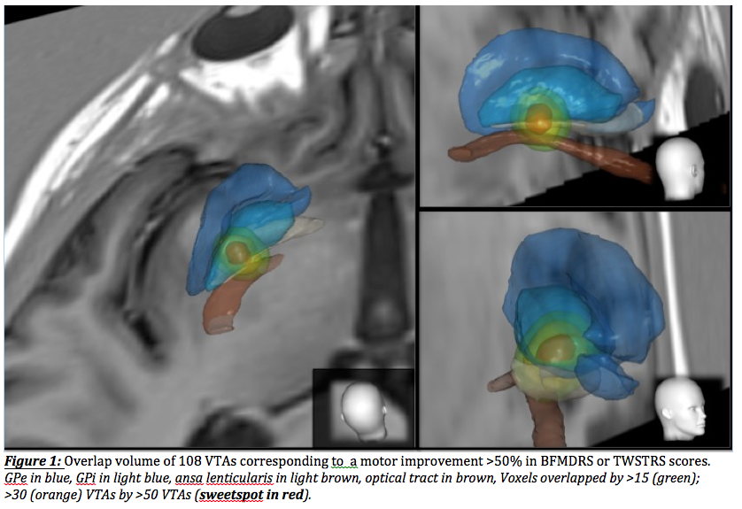
Methods: 85 subjects with dystonia (41 cervical mean TWSTRS 20.3±3.6 points/44 generalized dystonia, mean BFMDRS 45.8±20.5 points) under chronic bilateral GPi-DBS from 8 European DBS centres were stratified for chronic motor improvement (median reduction of 46.7(±27.7)% after 12.0 months in cervical / median reduction of 52.3(±35.9)% after 34.8 months in generalised dystonia). We simulated VTAs for each lead in subject’s related MRI space based on chronic stimulation parameters obtained from a chart review and associated with BFMDRS/TWSTRS improvement. All patient images were registered to a common average MRI. Only VTAs with a motor improvement >50% were taken for the visualisation of three different areas, defined by allegorizing only voxels that were overlapped by >15(green); >30(orange) VTAs and the “SweetSpot”, overlap volume of >50(red) VTAs.
Results: Wide variability of lead location, stimulation parameters and chronic motor improvement was observed in this cohort of 85 subjects. VTA size did not exhibit a significant correlation with improvement in motor symptoms. Model-based analysis of 108 responding VTAs showed a core mean volume (=”sweetspot”) located within and below the ventroposterior GPi. Stereotactic coordinates of the center of gravity were lateral:20.0, anterior:2.3 and inferior:2.6 (based on AC-PC in mm).
Conclusions: In this study, we showed that the magnitude of current injection is not decisive for the therapeutic DBS effect. In fact, the outcome is highly correlated with the precise location of neuromodulation within the region of interest. The most beneficial (sweet-)spot hints to a relevant contribution of subpallidal white matter, which could indicate a possible modulation of the ansa lenticularis for the anti-dystonic effect of DBS in addition to stimulation of the presumed sensorimotor region of the GPi
To cite this abstract in AMA style:
M. Reich, L. Lange, J. Roothans, A. Horn, F. Wodarg, J. Runge, M. Aström, F. Steigerwald, K. Witt, R. Nickl, P. Plettig, M. Wittstock, V. Coenen, P. Mahlknecht, W. Poewe, W. Eisner, C. Matthies, A. Kühn, J. Krauss, G. Deuschl*, J. Volkmann*. SweetSpot of antidystonic effect in pallidal neurostimulation: a European multicentre imaging study [abstract]. Mov Disord. 2017; 32 (suppl 2). https://www.mdsabstracts.org/abstract/sweetspot-of-antidystonic-effect-in-pallidal-neurostimulation-a-european-multicentre-imaging-study/. Accessed July 3, 2025.« Back to 2017 International Congress
MDS Abstracts - https://www.mdsabstracts.org/abstract/sweetspot-of-antidystonic-effect-in-pallidal-neurostimulation-a-european-multicentre-imaging-study/

